CHICAGO - Shear-wave elastography can be useful for evaluating tendons in patients with chronic midsubstance Achilles tendinopathy, suggests a study shown November 30 at the RSNA annual meeting.
In his talk, Dr. Scott Crawford, PhD, from the University of Wisconsin presented the results, which found lower shear-wave speed in the free tendon compared to the contralateral limb.
"Using shear-wave speeds to estimate tendon stiffness may be beneficial for clinical studies to address treatment effects on tendon remodeling," Crawford said.
Patients with Achilles tendinopathy typically present with pain and stiffness, resulting in difficulty walking. Pathology manifests via alterations in the microstructure composition of the symptomatic leg.
Ultrasound helps characterize some of these changes in these microstructures, but Crawford noted that traditional ultrasonic measures are limited by echogenicity issues and interrater reliability. Shear-wave elastography helps as an adjunctive tool by further assessing tendon properties, including tissue stiffness.
Previous reports suggest that elastography has high sensitivity in shear-wave speeds, but most of these are limited to the free tendon, Crawford said. Crawford et al wanted to expand on this by including multiple regions in the Achilles tendon. They compared quantitative measures of shear-wave speeds along the entire length of the tendon.
For their pilot study, they looked at data from 20 patients with an average of 56.3 years. The patients were asked to walk up and down a hallway for about six minutes to establish equilibrium in both legs before imaging took place. For ultrasonic imaging, the team had patients lie down in the prone position.
Researchers led by Dr. Scott Crawford from the University of Wisconsin found that shear-wave elastography is helpful in measuring stiffness of the free tendon in patients with chronic midsubstance Achilles tendinopathy. Image courtesy of Dr. Scott Crawford, PhD.The group found significant differences in shear-wave speed in the free tendon between the contralateral and symptomatic limb. The difference was 0.56 m/s, with the symptomatic leg showing slower speed (p = 0.03). Crawford noted that the differences in shear-wave speeds seen in the free tendon and healthy controls in this study were larger than that of the free tendon and contralateral limb as reported in previous research.
However, the researchers also found no differences between limbs in either the soleus aponeurosis (difference, 0.04; p = 0.13) or gastrocnemius aponeurosis (difference, -0.27; p = 0.99).
Crawford said expanding on these results in larger studies could position shear-wave speed as an adjunctive measure to not only characterize painful tendinopathy, but also help guide rehabilitation, intervention, and return to activity.




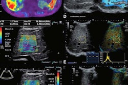
![Examples of ultrasound findings and techniques. (A) Images in a 39-year-old male patient with a mass in the left thigh. The mass is heterogeneous on the B-mode US image (compared with the patient in D) and showed increased microvascularity (superb microvascular imaging [SMI]) and shear-wave elastography (SWE) values. Undifferentiated pleomorphic sarcoma was diagnosed at biopsy (with pleomorphic rhabdomyosarcoma in surgical specimen). (B) Images in an 18-year-old male patient with a mass in the left leg. The mass is hypoechoic on the B-mode image, with no other findings suggestive of malignancy. The lesion is in contact with the cortex of the tibia, which is slightly irregular. CT revealed a doubtful anteromedial tibial erosion. The microvascular study demonstrated high vascularization, suggestive of malignancy. Periosteal Ewing sarcoma was diagnosed with both histologic and immunohistochemical confirmation. (C) Images in a 69-year-old female patient with a lump growing on the outside of the left leg. Multiple SWE examinations were performed (please note the high values obtained in the measurements, whereas the color map highlights the stiffness relative to adjacent tissues). SMI showed areas of increased vascularization to target for sampling. Undifferentiated spindle cell sarcoma was diagnosed at biopsy, with residual leiomyosarcoma in the surgical specimen after neoadjuvant therapy. (D) Images in a 56-year-old female patient with a mass in the right thigh. The mass is heterogeneous at both B-mode ultrasound (similar to patient A) and MRI (coronal T2-weighted spectral attenuated inversion recovery [SPAIR]; T1-weighted pre-contrast and postcontrast imaging), which even shows uptake after the administration of paramagnetic contrast material, which is traditionally suggestive of malignancy. Low values at SMI and elastography are suggestive of benignity. Spindle cell lipoma was diagnosed at biopsy, with atypical spindle cell lipomatous tumor in the surgical specimen.](https://img.auntminnie.com/mindful/smg/workspaces/default/uploads/2026/08/images-radiol250278fig2.APCFLSvX6p.jpg?auto=format%2Ccompress&fit=crop&h=100&q=70&w=100)


![Examples of ultrasound findings and techniques. (A) Images in a 39-year-old male patient with a mass in the left thigh. The mass is heterogeneous on the B-mode US image (compared with the patient in D) and showed increased microvascularity (superb microvascular imaging [SMI]) and shear-wave elastography (SWE) values. Undifferentiated pleomorphic sarcoma was diagnosed at biopsy (with pleomorphic rhabdomyosarcoma in surgical specimen). (B) Images in an 18-year-old male patient with a mass in the left leg. The mass is hypoechoic on the B-mode image, with no other findings suggestive of malignancy. The lesion is in contact with the cortex of the tibia, which is slightly irregular. CT revealed a doubtful anteromedial tibial erosion. The microvascular study demonstrated high vascularization, suggestive of malignancy. Periosteal Ewing sarcoma was diagnosed with both histologic and immunohistochemical confirmation. (C) Images in a 69-year-old female patient with a lump growing on the outside of the left leg. Multiple SWE examinations were performed (please note the high values obtained in the measurements, whereas the color map highlights the stiffness relative to adjacent tissues). SMI showed areas of increased vascularization to target for sampling. Undifferentiated spindle cell sarcoma was diagnosed at biopsy, with residual leiomyosarcoma in the surgical specimen after neoadjuvant therapy. (D) Images in a 56-year-old female patient with a mass in the right thigh. The mass is heterogeneous at both B-mode ultrasound (similar to patient A) and MRI (coronal T2-weighted spectral attenuated inversion recovery [SPAIR]; T1-weighted pre-contrast and postcontrast imaging), which even shows uptake after the administration of paramagnetic contrast material, which is traditionally suggestive of malignancy. Low values at SMI and elastography are suggestive of benignity. Spindle cell lipoma was diagnosed at biopsy, with atypical spindle cell lipomatous tumor in the surgical specimen.](https://img.auntminnie.com/mindful/smg/workspaces/default/uploads/2026/08/images-radiol250278fig2.APCFLSvX6p.jpg?auto=format%2Ccompress&fit=crop&h=112&q=70&w=112)