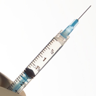
Adults taking corticosteroid injections for pain management that are guided by ultrasound and fluoroscopy have a lower incidence of symptomatic COVID-19, suggests research published July 5 in Radiology.
A team led by Dr. Joao Vicentini from Massachusetts General Hospital also found that study participants who had two or more visits for injections did not show a significantly higher infection rate than the general population. The group also found that participants diagnosed with COVID-19 had a higher body mass index (BMI).
"These findings provide reassurance to providers and individuals who rely on corticosteroid injections for the management of musculoskeletal pain during potential new COVID-19 surges, even in places with low vaccination rates," Vicentini and colleagues wrote.
Corticosteroid injections treat musculoskeletal pain, but they also temporarily suppress the immune system. This suppression can range between one day and four weeks, previous studies suggest.
This phenomenon was a cause for concern during the first months of the COVID-19 pandemic in the U.S. when it came to intra-articular and spinal injections. Professional societies suggested halting the use of injections or using alternative treatment methods. The study authors wrote that this led to disruptions in musculoskeletal treatments worldwide.
While previous research indicates that people who received injections during the lockdown period of the pandemic were relatively fine, the researchers wrote that results aren't well-known for the recovery period. This was when businesses started to reopen and physical distancing measures decreased but vaccines were not yet available.
Vicentini et al wanted to find out the incidence of symptomatic COVID-19 disease in people receiving corticosteroid injections guided by ultrasound and fluoroscopy and compare the results to the general population during the recovery period of the pandemic.
The team looked at prospective data from 2,714 corticosteroid injections performed between April 2020 and February 2021 for 2,190 adults with an average age of 59 years. Out of the total, 1,159 participants were men and 1,031 were women.
The researchers wrote that follow-up data were available for 1,960 (89%) participants who received 2,484 injections. Follow-up happened within a range of 28 days and 141 days after injections.
For the follow-up cohort, 10 (0.5%) people tested positive for COVID-19 within 28 days of receiving injections and 43 people tested positive up to four months after injection.
The team found that this was lower than the incidence rate in Massachusetts' general population, with 519,195 people testing positive for COVID-19 out of 6,892,503 (7.5%). The results were statistically significant for both 28-day and four-month marks (p < 0.001). The researchers also found that people in the study who were diagnosed with COVID-19 at 28 days (n = 10) had higher BMI, at 32, when compared with the entire study population, at 28 (p = 0.04).
The study authors wrote that "a few" different factors may have played a part in the lower incidence rates. These include study participants being screened for symptoms and close contacts prior to their injection, abiding by safety measures more, and possibly missing asymptomatic infection cases.



![Examples of ultrasound findings and techniques. (A) Images in a 39-year-old male patient with a mass in the left thigh. The mass is heterogeneous on the B-mode US image (compared with the patient in D) and showed increased microvascularity (superb microvascular imaging [SMI]) and shear-wave elastography (SWE) values. Undifferentiated pleomorphic sarcoma was diagnosed at biopsy (with pleomorphic rhabdomyosarcoma in surgical specimen). (B) Images in an 18-year-old male patient with a mass in the left leg. The mass is hypoechoic on the B-mode image, with no other findings suggestive of malignancy. The lesion is in contact with the cortex of the tibia, which is slightly irregular. CT revealed a doubtful anteromedial tibial erosion. The microvascular study demonstrated high vascularization, suggestive of malignancy. Periosteal Ewing sarcoma was diagnosed with both histologic and immunohistochemical confirmation. (C) Images in a 69-year-old female patient with a lump growing on the outside of the left leg. Multiple SWE examinations were performed (please note the high values obtained in the measurements, whereas the color map highlights the stiffness relative to adjacent tissues). SMI showed areas of increased vascularization to target for sampling. Undifferentiated spindle cell sarcoma was diagnosed at biopsy, with residual leiomyosarcoma in the surgical specimen after neoadjuvant therapy. (D) Images in a 56-year-old female patient with a mass in the right thigh. The mass is heterogeneous at both B-mode ultrasound (similar to patient A) and MRI (coronal T2-weighted spectral attenuated inversion recovery [SPAIR]; T1-weighted pre-contrast and postcontrast imaging), which even shows uptake after the administration of paramagnetic contrast material, which is traditionally suggestive of malignancy. Low values at SMI and elastography are suggestive of benignity. Spindle cell lipoma was diagnosed at biopsy, with atypical spindle cell lipomatous tumor in the surgical specimen.](https://img.auntminnie.com/mindful/smg/workspaces/default/uploads/2026/08/images-radiol250278fig2.APCFLSvX6p.jpg?auto=format%2Ccompress&fit=crop&h=100&q=70&w=100)


![Examples of ultrasound findings and techniques. (A) Images in a 39-year-old male patient with a mass in the left thigh. The mass is heterogeneous on the B-mode US image (compared with the patient in D) and showed increased microvascularity (superb microvascular imaging [SMI]) and shear-wave elastography (SWE) values. Undifferentiated pleomorphic sarcoma was diagnosed at biopsy (with pleomorphic rhabdomyosarcoma in surgical specimen). (B) Images in an 18-year-old male patient with a mass in the left leg. The mass is hypoechoic on the B-mode image, with no other findings suggestive of malignancy. The lesion is in contact with the cortex of the tibia, which is slightly irregular. CT revealed a doubtful anteromedial tibial erosion. The microvascular study demonstrated high vascularization, suggestive of malignancy. Periosteal Ewing sarcoma was diagnosed with both histologic and immunohistochemical confirmation. (C) Images in a 69-year-old female patient with a lump growing on the outside of the left leg. Multiple SWE examinations were performed (please note the high values obtained in the measurements, whereas the color map highlights the stiffness relative to adjacent tissues). SMI showed areas of increased vascularization to target for sampling. Undifferentiated spindle cell sarcoma was diagnosed at biopsy, with residual leiomyosarcoma in the surgical specimen after neoadjuvant therapy. (D) Images in a 56-year-old female patient with a mass in the right thigh. The mass is heterogeneous at both B-mode ultrasound (similar to patient A) and MRI (coronal T2-weighted spectral attenuated inversion recovery [SPAIR]; T1-weighted pre-contrast and postcontrast imaging), which even shows uptake after the administration of paramagnetic contrast material, which is traditionally suggestive of malignancy. Low values at SMI and elastography are suggestive of benignity. Spindle cell lipoma was diagnosed at biopsy, with atypical spindle cell lipomatous tumor in the surgical specimen.](https://img.auntminnie.com/mindful/smg/workspaces/default/uploads/2026/08/images-radiol250278fig2.APCFLSvX6p.jpg?auto=format%2Ccompress&fit=crop&h=112&q=70&w=112)