Corn flour, gelatin, olives, and raisins. Mix these and other ingredients together and you have yourself a nice ultrasound phantom model, at least according to research published July 28 in the British Journal of Radiology.
A team led by Dr. Elisabetta Giannotti from Nottingham City Hospital in the U.K. developed their phantom using household food ingredients and kitchenware to mimic soft tissue. Trainees showed highly positive responses to using it.
“We feel this phantom is realistic in terms of mimicking breast tissue,” said co-author Dr. Christopher Clarke from Queens Medical Centre in Nottingham to AuntMinnie.com. “It is approximately 10 pounds ($12.13) to make. Compared to other commercial phantoms, this is much cheaper, as commercial phantoms can be hundreds of dollars.”
Ultrasound is used for many interventional procedures for real-time visualization, including core needle biopsy, fluid aspiration, abscess drainage, and vascular access. Trainees need to practice for these procedures, which in many centers means performing on patients under direct supervision.
Simulation has been explored as an option for trainees to practice without the risk of harming patients, but commercially available ultrasound phantoms have high costs and may degrade with repeated use. Raw meat works well as a phantom, but the high bacteria levels present a health risk in clinical environments.
Giannotti et al wanted to develop a gelatin-based phantom with a corn flour additive, using materials found in a typical supermarket. These include the following:
- Hot water from the tap
- Corn flour
- Powdered gelatin (or vegan alternative)
- Food coloring (optional)
- Tofu
- Pitted olives
- Raisins
Overall, the phantom has a preparation time of 20 minutes, with a set time of 12 hours to solidify to the consistency of panna cotta, a creamy gelatin dessert.
The team also developed a cyst phantom made from a small water balloon or a finger from a disposable glove filled with water. They wrote that the phantom can be disposed of with other food waste. They added that the phantom will eventually fragment, typically after around 30 passes of the biopsy needle.
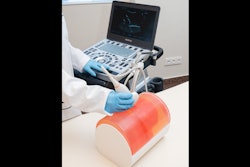
 Researchers in Nottingham, England developed an ultrasound phantom that they say mimics soft breast tissue and allows for training in all practical aspects of performing ultrasound-guided biopsy including needle-probe position via A) visualizing the target and B) visualizing the needle passing through the target during a biopsy. Structures inside the phantom include pitted olives stuffed with raisins and balloons representing cysts. Images courtesy of the BIR.
"This is sufficient to allow for training one to two people, and multiple phantoms can be prepared if required," the researchers wrote.
While the proposed phantom has been in use for about five years by the team, the study authors also wanted to find out what trainees think of making their own phantoms. Using a 1 to 5 score Likert scale, Giannotti and colleagues surveyed a total of 22 trainees to gather their thoughts.
| Opinions of radiology trainees on home-made ultrasound phantom model | |
| Survey question | Average score on Likert scale 1 (strongly disagree) to 5 (strongly agree) |
| "I found it easy to follow the instructions to create the ultrasound phantom biopsy model." | 4.5 |
| "The ultrasound phantom functioned well to demonstrate interventional techniques." | 4.7 |
The study authors wrote that the phantom's appearance on ultrasound images is very similar to that of human breast tissue. The size and shape of the phantom can also be varied depending on the containers used.
“One nice thing is when using on courses, everyone’s phantom is slightly different so we can practice on different coloured models with a varied array of targets and target locations,” Clarke said.
Clarke also told AuntMinnie.com that the positive response from students is multifactorial. This includes the trainees having some ownership of the model that they make themselves, as well as exposing trainees to ultrasound interventional simulation.
So, why these ingredients? The researchers wrote that a layer of tofu on top of the phantom mimics the skin and makes the phantom more resistant to fracturing during practice. The tofu "skin" also provides feedback with a similar consistency to human tissue. It can also hold a needle in place and not generate an obvious needle track.
The corn flour, meanwhile, provides "a truer reflection" of the in-vivo situation, the researchers wrote. It also gives the phantom an opaque look, hiding the biopsy needle target from external view.
The team also noted that for core biopsy practice, accurate targeting of the center of the "lesion" is confirmed via the presence of olive and raisin, with the raisins being stuffed inside the pitted olives. Also, inserting 3 ml to 5 ml of ultrasound gel into the phantom can mimic abscess drainage.
"Our gelatin phantom is low cost, hygienic, and uses readily available ingredients," the team concluded. "It is easy to produce and receives excellent trainee feedback and so provides an efficient and effective phantom for simulation training of ultrasound-guided procedures."