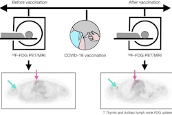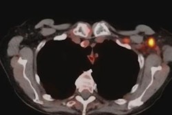
Unilateral axillary adenopathy after receiving the Pfizer-BioNTech COVID-19 vaccination can be visualized with ultrasound scanning, according to Spanish research published October 16 in European Radiology.
A team led by Dr. Alba Cristina Igual-Rouilleault from the University of Navarra Clinic in Spain found that ultrasound showed that people who had received the COVID-19 vaccine had a larger number of visible nodes, as well as nodes that had a greater maximum diameter and cortical thickness, among other characteristics.
"Both doses of COVID-19 vaccine induced an increase of all axillary lymph node parameters with statistically significant differences, especially in coronavirus-naïve patients," Igual-Rouilleault and colleagues wrote.
The COVID-19 vaccine has been documented in recent months as having side effects on patients such as pain, tenderness, or swelling at the site of vaccine administration, otherwise known as unilateral axillary lymphadenopathy.
This means radiologists should consider recent COVID-19 vaccination history as a possible differential diagnosis for patients with unilateral axillary adenopathy, such as during breast cancer screening, researchers said.
However, there is a lack of detailed information reporting gradual imaging of axillary nodes in vaccinated patients. The authors wrote that they wanted to assess the effects of the Pfzer-BioNTech vaccination on unilateral axillary lymph nodes, comparing nodal basal features with their characteristics after the first and second vaccine doses.
The researchers looked at 91 volunteer employees from the university clinic who participated in the Pfizer-BioNTech vaccination campaign. The volunteers were between the ages of 20 and 65, with an average age of 44 years. Of the total, 72 were females, and a previous history of COVID-19 infection was registered in 26 patients.
Three axillary ultrasound evaluations of the ipsilateral vaccinated arm were performed. They were done before vaccination (baseline), the week after the first dose, and the week after the second dose.
The team recorded the following characteristics: the total number of visible nodes, the maximum measurements of the diameter and cortex, Bedi's classification, and color Doppler evaluation.
| Characteristics on ultrasound of patients with enlarged nodes before and after COVID-19 vaccination | |||
| Baseline | 1st follow-up | 2nd follow-up | |
| No. of Nodes (average) | 2.96 | 4.77 | 6.21 |
| Diameter (average) | 15.8 mm | 21.1 mm | 24.0 mm |
| Cortex (average) | 1.6 mm | 3.7 mm | 4.6 mm |
The researchers found that patients who were not previously infected by SARS-CoV-2 showed a more significant lymph node response compared to those who were previously infected. They also found that axillary nodes showed benign characteristics more frequently, with mild cortical thickness and preserved uniformity.
However, the study authors said that malignancy exclusion is required in oncologic follow-ups or patients recently diagnosed with breast cancer. They also said percutaneous ultrasound-guided needle biopsy or a short-term follow-up ultrasound is necessary.
More than 50% of patients normalized their cortex value the month after the second vaccination. The team wrote that more studies should be performed to determine the time required to reach basal cortical values.
Igual-Rouilleault and colleagues said this study may be used to help properly manage ultrasound axillary lymphadenopathy in the era of COVID-19 vaccination by identifying subgroups of patients that need close monitoring and perhaps for developing guidelines for clinical use.
"However, further research should be performed to understand and decipher underlying mechanisms," they added.


![Examples of ultrasound findings and techniques. (A) Images in a 39-year-old male patient with a mass in the left thigh. The mass is heterogeneous on the B-mode US image (compared with the patient in D) and showed increased microvascularity (superb microvascular imaging [SMI]) and shear-wave elastography (SWE) values. Undifferentiated pleomorphic sarcoma was diagnosed at biopsy (with pleomorphic rhabdomyosarcoma in surgical specimen). (B) Images in an 18-year-old male patient with a mass in the left leg. The mass is hypoechoic on the B-mode image, with no other findings suggestive of malignancy. The lesion is in contact with the cortex of the tibia, which is slightly irregular. CT revealed a doubtful anteromedial tibial erosion. The microvascular study demonstrated high vascularization, suggestive of malignancy. Periosteal Ewing sarcoma was diagnosed with both histologic and immunohistochemical confirmation. (C) Images in a 69-year-old female patient with a lump growing on the outside of the left leg. Multiple SWE examinations were performed (please note the high values obtained in the measurements, whereas the color map highlights the stiffness relative to adjacent tissues). SMI showed areas of increased vascularization to target for sampling. Undifferentiated spindle cell sarcoma was diagnosed at biopsy, with residual leiomyosarcoma in the surgical specimen after neoadjuvant therapy. (D) Images in a 56-year-old female patient with a mass in the right thigh. The mass is heterogeneous at both B-mode ultrasound (similar to patient A) and MRI (coronal T2-weighted spectral attenuated inversion recovery [SPAIR]; T1-weighted pre-contrast and postcontrast imaging), which even shows uptake after the administration of paramagnetic contrast material, which is traditionally suggestive of malignancy. Low values at SMI and elastography are suggestive of benignity. Spindle cell lipoma was diagnosed at biopsy, with atypical spindle cell lipomatous tumor in the surgical specimen.](https://img.auntminnie.com/mindful/smg/workspaces/default/uploads/2026/08/images-radiol250278fig2.APCFLSvX6p.jpg?auto=format%2Ccompress&fit=crop&h=100&q=70&w=100)


![Examples of ultrasound findings and techniques. (A) Images in a 39-year-old male patient with a mass in the left thigh. The mass is heterogeneous on the B-mode US image (compared with the patient in D) and showed increased microvascularity (superb microvascular imaging [SMI]) and shear-wave elastography (SWE) values. Undifferentiated pleomorphic sarcoma was diagnosed at biopsy (with pleomorphic rhabdomyosarcoma in surgical specimen). (B) Images in an 18-year-old male patient with a mass in the left leg. The mass is hypoechoic on the B-mode image, with no other findings suggestive of malignancy. The lesion is in contact with the cortex of the tibia, which is slightly irregular. CT revealed a doubtful anteromedial tibial erosion. The microvascular study demonstrated high vascularization, suggestive of malignancy. Periosteal Ewing sarcoma was diagnosed with both histologic and immunohistochemical confirmation. (C) Images in a 69-year-old female patient with a lump growing on the outside of the left leg. Multiple SWE examinations were performed (please note the high values obtained in the measurements, whereas the color map highlights the stiffness relative to adjacent tissues). SMI showed areas of increased vascularization to target for sampling. Undifferentiated spindle cell sarcoma was diagnosed at biopsy, with residual leiomyosarcoma in the surgical specimen after neoadjuvant therapy. (D) Images in a 56-year-old female patient with a mass in the right thigh. The mass is heterogeneous at both B-mode ultrasound (similar to patient A) and MRI (coronal T2-weighted spectral attenuated inversion recovery [SPAIR]; T1-weighted pre-contrast and postcontrast imaging), which even shows uptake after the administration of paramagnetic contrast material, which is traditionally suggestive of malignancy. Low values at SMI and elastography are suggestive of benignity. Spindle cell lipoma was diagnosed at biopsy, with atypical spindle cell lipomatous tumor in the surgical specimen.](https://img.auntminnie.com/mindful/smg/workspaces/default/uploads/2026/08/images-radiol250278fig2.APCFLSvX6p.jpg?auto=format%2Ccompress&fit=crop&h=112&q=70&w=112)