PET/MRI scans have revealed immune system reactions after COVID-19 vaccines in children with cancer, according to a study published October 26 in the Journal of Nuclear Medicine.
A team at Stanford University in Stanford, CA, evaluated F-18 FDG-PET/MRI scans of six children and young adults before and after COVID-19 vaccination. The scans demonstrated thymus activation in addition to local lymph node reactions, the group found.
“Understanding postvaccination imaging findings in patients with cancer is important because these results may be confounded with tumor relapse or metastasis,” wrote first author Gaurav Luthria, MD, PhD, and colleagues.
In patients with cancer, an increase in the size and metabolic activity of lymph nodes can indicate tumor recurrence or progression and play an important role in guiding treatment decisions, the author explained.
Several studies have demonstrated that vaccine-related lymphadenopathy may confound disease assessment in oncology patients, the researchers added. In this study, they hypothesized that F-18 FDG-PET/MRI may reveal activation in the thymus in pediatric oncology patients in addition to lymph node reactions.
The group assessed scans of six children with extrathoracic cancer before and after COVID-19 vaccination. They analyzed both PET and MRI biomarkers, such as uptake of F-18 FDG radiotracer (maximum standard uptake values, or SUVmax), mean apparent diffusion coefficient on MRI, and size of the thymus and axillary lymph nodes.
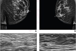
There was a significant increase in the metabolic activity in the ipsilateral axillary lymph nodes across all patients after COVID-19 vaccination, according to the findings. The mean SUVmax increased by a factor of three, from 0.87 before vaccination to 2.61 after vaccination (p = 0.03).
 A graphic abstract describing the study. Image courtesy of the Journal of Nuclear Medicine.
In addition, all six patients demonstrated an increased size, increased metabolic activity, and restricted diffusion of the lymph nodes and thymus after COVID-19 vaccination. The mean SUVmax of the thymus increased twofold, from 1.77 before vaccination to 3.46 after vaccination.
Finally, they noted that the mean size of the thymus, approximated as the product of the shortest and longest transverse diameters, increased 1.7-fold, from 7.02 centimeters to 11.91 centimeters.
“Here we show for the first time, to our knowledge, that F-18 FDG-PET/MRI can reveal thymus activation in addition to local lymph node reactions in children after COVID-19 vaccination,” the authors wrote.
Ultimately, the study underscores the importance of verifying a patient’s vaccination status before an imaging examination, as vaccination-associated changes can lead to false-positive diagnoses, the authors noted.
“The detection of increased F-18 FDG activity in the local lymph nodes and thymus after COVID-19 vaccination on F-18 FDG-PET/MRI scans could be helpful to confirm a vaccine-induced immune response in cancer patients,” the researchers concluded.
The full article is available here.