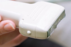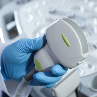
The answer to preventing cross-contamination from repeat use of ultrasound probes may not come in the form of extensive cleaning or antibacterial chemicals but rather a simple strip of adhesive tape.
That's what a team led by Yuan Qin, PhD, from the Beijing University of Chemical Technology found in a study published October 6 in Surfaces and Interfaces.
The researchers say using acrylate pressure-sensitive adhesive (PSA) tape as an ultrasound probe cover not only prevents cross-infection but also has less impact on the clarity of ultrasound images and saves time for radiologists.
"The ease of preparation, convenience of use, and effectiveness of acrylate PSA tapes indicate that they have broad application prospects for sonographic examinations," Qin and colleagues wrote.
Ultrasound probes are an essential part of medical ultrasound imaging. The most commonly used cleaning methods include using paper towels to wipe out coupling gel and chemically treating probes with alcohol or quaternary ammonium compounds. However, researchers said damages to the probes and cross-infection between patients might be unavoidable.
Radiologists should also eliminate ultrasound transmission gel on the probes after scanning. However, researchers said this is a time-consuming process.
"Nowadays, how to improve convenience and how to reduce the time between examinations are important issues to be considered because ultrasound diagnosis is required quickly and effectively in the face of many patients," Qin et al wrote.
Acrylate pressure-sensitive adhesive tape is already used for medical applications such as wound coverings and transdermal drug delivery systems. The researchers touted its nontoxic, transparent, and mechanical properties, as well as its tendency to leave no residue behind.
The team wanted to test the effectiveness and feasibility of pressure-sensitive adhesive tape on preventing cross-contamination and improving productivity.
Probes were used on a skin-like silicone puncture phantom that simulated human tissue. Three types of glue were used for the tapes, and air bubbles were rolled out as the tapes were added to the probes. The glues include Polyethylene terephthalate, polyurethane, and polyethylene.
Polyethylene film with good softness, little effect on the clarity of ultrasonic images, and substrate versatility, was selected as the underlying layer of film for the tapes.
Results showed that the shear strength of tapes improved with increasing molecular weight of acrylate copolymer. However, tack and 180° peel strength showed a downward trend.
Forming a cross-linked network among polymer chains also greatly improved the shear strength of tapes to meet the requirements of ultrasound probe covers, including being easy to peel from the ultrasound probe, easy to adhere to the probe, not leaving residue behind, and having less impact on the clarity of ultrasound images.
The researchers also found that a viscoelastic adhesive layer of tape can replace the coupling gel between ultrasound probe and pressure-sensitive adhesive tape.
A clinician judged the quality of images taken with the taped probes and said tape containing polyethylene and polyurethane had "excellent" image clarity, as well as "excellent" softness for contact with human skin.
The study authors said that compared with conventional disinfecting procedures, pressure-sensitive adhesive tape for probes has three advantages. They included preventing direct contact with the human body to reduce cross-infection and lens degradation, minimizing downtime between patients through easy tape adherence and removal, and replacing the coupling gel, which could help avoid paper towel usage and minimize cross-infection risk.
They added that it could also reduce the occurrence probability of corrosion and electric leakage.
"[The tape] is expected to find applications in sonographic examinations in the future to protect ultrasound probes and to prevent cross-infections," Qin and colleagues wrote.


![Examples of ultrasound findings and techniques. (A) Images in a 39-year-old male patient with a mass in the left thigh. The mass is heterogeneous on the B-mode US image (compared with the patient in D) and showed increased microvascularity (superb microvascular imaging [SMI]) and shear-wave elastography (SWE) values. Undifferentiated pleomorphic sarcoma was diagnosed at biopsy (with pleomorphic rhabdomyosarcoma in surgical specimen). (B) Images in an 18-year-old male patient with a mass in the left leg. The mass is hypoechoic on the B-mode image, with no other findings suggestive of malignancy. The lesion is in contact with the cortex of the tibia, which is slightly irregular. CT revealed a doubtful anteromedial tibial erosion. The microvascular study demonstrated high vascularization, suggestive of malignancy. Periosteal Ewing sarcoma was diagnosed with both histologic and immunohistochemical confirmation. (C) Images in a 69-year-old female patient with a lump growing on the outside of the left leg. Multiple SWE examinations were performed (please note the high values obtained in the measurements, whereas the color map highlights the stiffness relative to adjacent tissues). SMI showed areas of increased vascularization to target for sampling. Undifferentiated spindle cell sarcoma was diagnosed at biopsy, with residual leiomyosarcoma in the surgical specimen after neoadjuvant therapy. (D) Images in a 56-year-old female patient with a mass in the right thigh. The mass is heterogeneous at both B-mode ultrasound (similar to patient A) and MRI (coronal T2-weighted spectral attenuated inversion recovery [SPAIR]; T1-weighted pre-contrast and postcontrast imaging), which even shows uptake after the administration of paramagnetic contrast material, which is traditionally suggestive of malignancy. Low values at SMI and elastography are suggestive of benignity. Spindle cell lipoma was diagnosed at biopsy, with atypical spindle cell lipomatous tumor in the surgical specimen.](https://img.auntminnie.com/mindful/smg/workspaces/default/uploads/2026/08/images-radiol250278fig2.APCFLSvX6p.jpg?auto=format%2Ccompress&fit=crop&h=100&q=70&w=100)


![Examples of ultrasound findings and techniques. (A) Images in a 39-year-old male patient with a mass in the left thigh. The mass is heterogeneous on the B-mode US image (compared with the patient in D) and showed increased microvascularity (superb microvascular imaging [SMI]) and shear-wave elastography (SWE) values. Undifferentiated pleomorphic sarcoma was diagnosed at biopsy (with pleomorphic rhabdomyosarcoma in surgical specimen). (B) Images in an 18-year-old male patient with a mass in the left leg. The mass is hypoechoic on the B-mode image, with no other findings suggestive of malignancy. The lesion is in contact with the cortex of the tibia, which is slightly irregular. CT revealed a doubtful anteromedial tibial erosion. The microvascular study demonstrated high vascularization, suggestive of malignancy. Periosteal Ewing sarcoma was diagnosed with both histologic and immunohistochemical confirmation. (C) Images in a 69-year-old female patient with a lump growing on the outside of the left leg. Multiple SWE examinations were performed (please note the high values obtained in the measurements, whereas the color map highlights the stiffness relative to adjacent tissues). SMI showed areas of increased vascularization to target for sampling. Undifferentiated spindle cell sarcoma was diagnosed at biopsy, with residual leiomyosarcoma in the surgical specimen after neoadjuvant therapy. (D) Images in a 56-year-old female patient with a mass in the right thigh. The mass is heterogeneous at both B-mode ultrasound (similar to patient A) and MRI (coronal T2-weighted spectral attenuated inversion recovery [SPAIR]; T1-weighted pre-contrast and postcontrast imaging), which even shows uptake after the administration of paramagnetic contrast material, which is traditionally suggestive of malignancy. Low values at SMI and elastography are suggestive of benignity. Spindle cell lipoma was diagnosed at biopsy, with atypical spindle cell lipomatous tumor in the surgical specimen.](https://img.auntminnie.com/mindful/smg/workspaces/default/uploads/2026/08/images-radiol250278fig2.APCFLSvX6p.jpg?auto=format%2Ccompress&fit=crop&h=112&q=70&w=112)