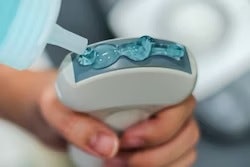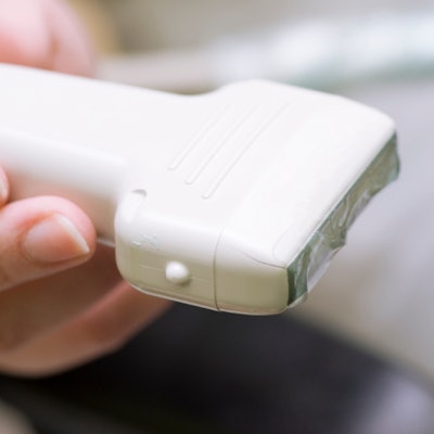
Point-of-care ultrasound (POCUS) needs rigorously designed trials to prove its value as the technology's popularity continues to grow among physicians, according to an editorial published October 11 in JAMA Internal Medicine.
In their editorial, Dr. Eden Bernstein from the Cleveland Clinic Foundation in Ohio and Dr. Tracy Wang from Duke University in North Carolina wrote that the use of POCUS for diagnostic purposes is not as firmly grounded in evidence and has "many unanswered questions" regarding its use.
"We recommend making POCUS use more evidence-based by focusing research efforts on assessing the role of POCUS in patient treatment and clinical outcomes," they said.
POCUS has emerged in recent years as a preferred choice in examining patients at bedside or even outside the clinical setting, with previous studies citing its ease of use, affordability, high sensitivity in some areas, and shorter diagnosis time. It has been met with so much enthusiasm by clinicians and radiologists that the American College of Physicians and the Society of Hospital Medicine have endorsed its use.
However, Bernstein and Wang wrote that there is a lack of formal data available when it comes to understanding the clinical benefit of POCUS. They also said images may not be easily accessed for quality assurance and retrospective analysis.
"Very few trials have examined meaningful clinical outcomes related to POCUS or even indirect clinical outcomes, such as time to treatment initiation, avoidance of transfer to a higher level of care, or length of stay," they said.
The two cited a 2018 study published in the Annals of Emergency Medicine that evaluated the ability of POCUS to improve clinical outcomes in 270 emergency department patients with undifferentiated hypotension. In the randomized clinical trial, the authors of the study did not find any benefits for survival, length of stay, rates of downstream CT scanning, inotropic use, or fluid administration.
"The addition of a point-of-care ultrasonography protocol to standard care may not translate into a survival benefit in this group," the researchers wrote.
Bernstein and Wang also noted the "virtually nonexistent" data on downstream adverse consequences of POCUS.
"We do not know the degree to which false-positive findings occur in practice and how often they lead to unnecessary testing or treatments that potentially worsen patient outcomes," they said.
The authors also wrote about training and education among POCUS users, saying there are no universally recognized standards for accreditation or credentialing. POCUS does not need to be performed by imaging experts.
Bernstein and Wang called for standard training to include education on when a POCUS examination could help guide management based on perceived pretest probability. They also said training should include technical skills for image gathering and interpretation targeted to non-imaging specialists.
"There is no doubt that POCUS use is viscerally satisfying and appeals to the clinician taking care of patients whose bedside history and physical examination cannot convey a clear diagnosis," they said. "Studies to date have largely been aimed at establishing the diagnostic utility of POCUS."