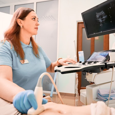
About two-thirds of sonographers report performing ultrasound scans while experiencing musculoskeletal pain, according to a survey study published September 17 in the Journal of Diagnostic Medical Sonography.
That said, the levels of pain reported in the new survey actually represent a decline compared with previous studies, according to Amanda Hogan from Bellin College in Wisconsin. Hogan found that while reported pain in sonographers is decreasing, certain specialties within sonography such as vascular, cardiac, and obstetrics/gynecology are more prone to injury and higher pain levels.
"The trend must continue to decrease pain and injury rates within the profession and improve overall ergonomics to keep decreasing the number of sonographers scanning in pain," Hogan said.
Sonographers struggle with the physical demands of their profession daily and face increased risk of musculoskeletal disorders as a result. Previous research has found between 75% and 90% of sonographers report scanning in pain. However, many sonographers are fearful of reporting their injuries to management.
"Sonographers historically felt that injury was part of their job," Hogan wrote. "A similar thought process still prevails today, with 57% of injured sonographers not reporting their injury to management."
The cost of work-related musculoskeletal disorders, meanwhile, is about $20 billion in direct worker's compensation costs and another estimated $100 billion in indirect costs such as lost revenue, training, and replacement, Hogan wrote. Previous research also suggests the cost of lost revenue due to sonographer injury to be $40,000 to $96,000 monthly.
Hogan wanted to find out if there is a difference in pain levels among sonographers in different specialties, as well as if certain specialties within sonography have higher injury rates.
The survey questionnaire was completed by 7,993 sonographers, 7,054 (88%) being female and 882 being male (11%). Pain levels were on a 1 to 10 scale with 10 being the highest.
The percentage of sonographers who reported scanning in pain across all specialties was 66.3%, with an average pain level of 4 out of 10. Sonographers scanning greater than 18 scans per day or greater than 30 minutes had the highest levels of perceived pain.
Males reported their primary specialty as being cardiac, abdominal, and vascular, while females selected ob/gyn and abdominal specialties.
A statistically significant number of male sonographers practiced in vascular and cardiac specialties compared with their female counterparts. More males (44%) reported not scanning in pain compared with 32.4% of females (p < 0.05).
The average pain level reported for males was 3.51 compared with 4.32 reported by females. Males reported a statistically significant lower level of pain in the shoulder, neck, and low back compared with female sonographers (p < 0.05).
Sonographers specializing in the cardiac, vascular, and ob/gyn specialties reported the highest pain and injury levels.
Shoulder pain was reported for all specialties, ranging from 63.9% for vascular to 69.7% for ob/gyn. Low back pain meanwhile was more associated with vascular specialists at 40.1%.
For ob/gyn sonographers, 51.5% reported pain in the hand and wrist, as well as a significantly lower amount of low back pain (28.5%) compared with all other specialties.
While no significant differences in pain level was found between ages, sonographers above the age of 50 reported a statistically significant increase in sustaining a work-related musculoskeletal disorder (p < 0.05).
Hogan said that the 66% figure reported in her study is lower than the 75% to 90% found in previous research. This could be due to differences in the survey sample of her study, but it could also indicate the effectiveness of interventions employed to raise awareness of ergonomic issues in sonography.
"Much work has been done by the sonography community to reduce pain and injury rates, but more attention is needed to address specialty-specific ergonomic needs and interventions," Hogan said. "Although this research that less sonographers are scanning in pain is promising, there is still 66% of sonographers who did report scanning in pain."


![Examples of ultrasound findings and techniques. (A) Images in a 39-year-old male patient with a mass in the left thigh. The mass is heterogeneous on the B-mode US image (compared with the patient in D) and showed increased microvascularity (superb microvascular imaging [SMI]) and shear-wave elastography (SWE) values. Undifferentiated pleomorphic sarcoma was diagnosed at biopsy (with pleomorphic rhabdomyosarcoma in surgical specimen). (B) Images in an 18-year-old male patient with a mass in the left leg. The mass is hypoechoic on the B-mode image, with no other findings suggestive of malignancy. The lesion is in contact with the cortex of the tibia, which is slightly irregular. CT revealed a doubtful anteromedial tibial erosion. The microvascular study demonstrated high vascularization, suggestive of malignancy. Periosteal Ewing sarcoma was diagnosed with both histologic and immunohistochemical confirmation. (C) Images in a 69-year-old female patient with a lump growing on the outside of the left leg. Multiple SWE examinations were performed (please note the high values obtained in the measurements, whereas the color map highlights the stiffness relative to adjacent tissues). SMI showed areas of increased vascularization to target for sampling. Undifferentiated spindle cell sarcoma was diagnosed at biopsy, with residual leiomyosarcoma in the surgical specimen after neoadjuvant therapy. (D) Images in a 56-year-old female patient with a mass in the right thigh. The mass is heterogeneous at both B-mode ultrasound (similar to patient A) and MRI (coronal T2-weighted spectral attenuated inversion recovery [SPAIR]; T1-weighted pre-contrast and postcontrast imaging), which even shows uptake after the administration of paramagnetic contrast material, which is traditionally suggestive of malignancy. Low values at SMI and elastography are suggestive of benignity. Spindle cell lipoma was diagnosed at biopsy, with atypical spindle cell lipomatous tumor in the surgical specimen.](https://img.auntminnie.com/mindful/smg/workspaces/default/uploads/2026/08/images-radiol250278fig2.APCFLSvX6p.jpg?auto=format%2Ccompress&fit=crop&h=100&q=70&w=100)


![Examples of ultrasound findings and techniques. (A) Images in a 39-year-old male patient with a mass in the left thigh. The mass is heterogeneous on the B-mode US image (compared with the patient in D) and showed increased microvascularity (superb microvascular imaging [SMI]) and shear-wave elastography (SWE) values. Undifferentiated pleomorphic sarcoma was diagnosed at biopsy (with pleomorphic rhabdomyosarcoma in surgical specimen). (B) Images in an 18-year-old male patient with a mass in the left leg. The mass is hypoechoic on the B-mode image, with no other findings suggestive of malignancy. The lesion is in contact with the cortex of the tibia, which is slightly irregular. CT revealed a doubtful anteromedial tibial erosion. The microvascular study demonstrated high vascularization, suggestive of malignancy. Periosteal Ewing sarcoma was diagnosed with both histologic and immunohistochemical confirmation. (C) Images in a 69-year-old female patient with a lump growing on the outside of the left leg. Multiple SWE examinations were performed (please note the high values obtained in the measurements, whereas the color map highlights the stiffness relative to adjacent tissues). SMI showed areas of increased vascularization to target for sampling. Undifferentiated spindle cell sarcoma was diagnosed at biopsy, with residual leiomyosarcoma in the surgical specimen after neoadjuvant therapy. (D) Images in a 56-year-old female patient with a mass in the right thigh. The mass is heterogeneous at both B-mode ultrasound (similar to patient A) and MRI (coronal T2-weighted spectral attenuated inversion recovery [SPAIR]; T1-weighted pre-contrast and postcontrast imaging), which even shows uptake after the administration of paramagnetic contrast material, which is traditionally suggestive of malignancy. Low values at SMI and elastography are suggestive of benignity. Spindle cell lipoma was diagnosed at biopsy, with atypical spindle cell lipomatous tumor in the surgical specimen.](https://img.auntminnie.com/mindful/smg/workspaces/default/uploads/2026/08/images-radiol250278fig2.APCFLSvX6p.jpg?auto=format%2Ccompress&fit=crop&h=112&q=70&w=112)