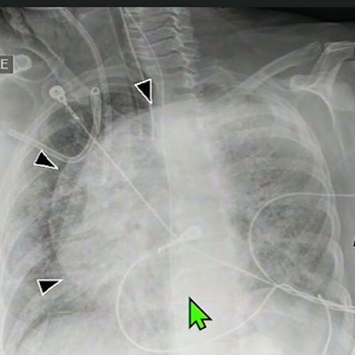
Portable chest x-rays acquired in critically ill patients in the prone position are comparable to those taken with patients in supine positions, according to a study presented May 3 at the American Roentgen Ray Society (ARRS) meeting in New Orleans.
Studies suggest that imaging patients with acute respiratory distress syndrome (ARDS) in prone positions is associated with a reduction in mortality, due to improved oxygen saturation in the lungs. Yet a paucity of literature exists regarding chest radiographs of prone patients, said Dr. Ami Rubinowitz, an associate professor of radiology and biomedical imaging at Yale School of Medicine in New Haven, CT.
"We don't know how well we can see the various support devices that these patients frequently have. We kind of all got slammed into it during COVID when proning of patients became commonplace," she said.
In the study presented at ARRS 2022, Rubinowitz and colleagues set out to look at image quality and distinguishing features of prone radiographs compared with images of patients in supine positions. They obtained 162 portable x-ray images from 11 hospitals performed between March 2020 and February 2021.

Critically ill patients underwent both supine and prone x-ray in the same day. Each set of images (81 each) was retrospectively reviewed for adequate imaging of the lungs by at least two cardiothoracic radiologists.
The reviewers found prone and supine positions were comparable for adequate imaging of the lung, with the base adequately imaged in 84% of prone images and 90% of supine images. The apex was adequately imaged in 93% of prone x-rays compared with 94% of supine x-rays, according to the findings.
However, analysis revealed the reviewers found clear position-dependent differences between prone and supine portable chest x-rays, as follows:
- Scapular tip locations beyond the rib border were greater in prone position images
- Stomach gas bubble in the fundus was greater in prone positioning images
- Displacement of one or both breast shadows in women were greater in prone position images
"Both techniques reliably assessed line and tube positioning," Rubinowitz added.
Ultimately, radiologists should take into account any changes in position when comparing to prior exams, and critical care teams especially can be assured that images of patients in prone position "do not diminish the ability to verify line placement," Rubinowitz concluded.