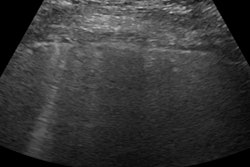
Point-of-care ultrasound (POCUS) performed better than chest x-ray in diagnosing atypical pneumonia that's presumed to be COVID-19, according to a study presented at the American College of Emergency Physicians (ACEP) annual meeting in late October.
Point-of-care ultrasound had higher sensitivity than chest x-ray for the task, although both modalities fell short in terms of specificity in comparison to CT. Researchers analyzed imaging for diagnosing COVID-19 cases in the early part of the pandemic, when access to polymerase chain reaction (PCR) tests was inconsistent.
"Many places were faced with, especially at the beginning of the pandemic, not having access to the PCR (nasopharyngeal) swabs, with trying to make a diagnosis (of atypical pneumonia/COVID-19) through alternative methods," explained Dr. Kendra Mendez, the study's presenting author and an ultrasound fellow at the Lewis Katz School of Medicine at Temple University in Philadelphia.
Mendez noted the data were collected prospectively over a two-week period in April at Temple University Hospital, which has more than 105,000 visits to the emergency department annually, as it experienced a surge during the pandemic.
"We already use lung ultrasound to differentiate between various conditions, differentiating between heart failure and [chronic obstructive pulmonary disease] exacerbation, for example," Mendez said. "The protocol of using lung ultrasound when someone is in respiratory distress is already in our department. We wanted to see the utility of it with COVID-19."
Some of the advantages of point-of-care ultrasound include portability and decreased burden with respect to infection control, according to Mendez.
"The portable units are much easier to disinfect than our larger machines," she said.
Mendez and co-investigators set up a protocol based on previous research of the appearance of atypical viral pneumonia on ultrasound or x-ray and used it to examine different areas of the lungs, comparing sensitivity and specificity of ultrasound versus chest x-ray. They used noncontrast CT as the gold standard.
They used an eight-zone lung ultrasound protocol and categorized predefined abnormalities as subpleural consolidations, irregular pleural lines, or pleural effusion.
Investigators enrolled 143 patients with signs and symptoms of COVID-19. A total of 70 had both a positive lung ultrasound and positive chest x-ray. Of those, 58 had positive CT findings suggestive of atypical viral pneumonia.
A total of 42 patients had a positive lung ultrasound and negative chest x-ray. Of those, 23 had positive CT findings. Six patients had a negative lung ultrasound and a positive chest x-ray, but none of the six had a positive CT finding. Twenty-five patients had both a negative lung ultrasound and negative chest x-ray. Still, seven of them advanced to CT imaging because they had one or more high-risk features, such as the presence of immunosuppression or lymphopenia. Two of the seven had a positive CT.
Lung ultrasound recorded higher sensitivity than chest x-ray, the researchers found.
| Chest x-ray vs. lung ultrasound for COVID-19 detection | ||
| Chest x-ray | Lung ultrasound | |
| Sensitivity | 69.9% | 97.6% |
| Specificity | 44.4% | 33.3% |
"Lung ultrasound is more sensitive than chest x-ray," said Dr. Mendez. "Both modalities had poor specificity."
The data's generalizability is limited by several variables, such as the fact that the study was performed at a single center, the disease prevalence was 75% in the study population, and the potential lack of familiarity with point-of-care ultrasound in a given emergency department, according to Mendez.
"All emergency medicine physicians in our institution are well-versed in point-of-care ultrasound, possibly limiting the utility (of point-of-care ultrasound) in the broader emergency medicine community," said Dr. Mendez.
Still, point-of-care ultrasound can be a triage tool, particularly in under-resourced settings, Mendez pointed out.

![Examples of ultrasound findings and techniques. (A) Images in a 39-year-old male patient with a mass in the left thigh. The mass is heterogeneous on the B-mode US image (compared with the patient in D) and showed increased microvascularity (superb microvascular imaging [SMI]) and shear-wave elastography (SWE) values. Undifferentiated pleomorphic sarcoma was diagnosed at biopsy (with pleomorphic rhabdomyosarcoma in surgical specimen). (B) Images in an 18-year-old male patient with a mass in the left leg. The mass is hypoechoic on the B-mode image, with no other findings suggestive of malignancy. The lesion is in contact with the cortex of the tibia, which is slightly irregular. CT revealed a doubtful anteromedial tibial erosion. The microvascular study demonstrated high vascularization, suggestive of malignancy. Periosteal Ewing sarcoma was diagnosed with both histologic and immunohistochemical confirmation. (C) Images in a 69-year-old female patient with a lump growing on the outside of the left leg. Multiple SWE examinations were performed (please note the high values obtained in the measurements, whereas the color map highlights the stiffness relative to adjacent tissues). SMI showed areas of increased vascularization to target for sampling. Undifferentiated spindle cell sarcoma was diagnosed at biopsy, with residual leiomyosarcoma in the surgical specimen after neoadjuvant therapy. (D) Images in a 56-year-old female patient with a mass in the right thigh. The mass is heterogeneous at both B-mode ultrasound (similar to patient A) and MRI (coronal T2-weighted spectral attenuated inversion recovery [SPAIR]; T1-weighted pre-contrast and postcontrast imaging), which even shows uptake after the administration of paramagnetic contrast material, which is traditionally suggestive of malignancy. Low values at SMI and elastography are suggestive of benignity. Spindle cell lipoma was diagnosed at biopsy, with atypical spindle cell lipomatous tumor in the surgical specimen.](https://img.auntminnie.com/mindful/smg/workspaces/default/uploads/2026/08/images-radiol250278fig2.APCFLSvX6p.jpg?auto=format%2Ccompress&fit=crop&h=100&q=70&w=100)


![Examples of ultrasound findings and techniques. (A) Images in a 39-year-old male patient with a mass in the left thigh. The mass is heterogeneous on the B-mode US image (compared with the patient in D) and showed increased microvascularity (superb microvascular imaging [SMI]) and shear-wave elastography (SWE) values. Undifferentiated pleomorphic sarcoma was diagnosed at biopsy (with pleomorphic rhabdomyosarcoma in surgical specimen). (B) Images in an 18-year-old male patient with a mass in the left leg. The mass is hypoechoic on the B-mode image, with no other findings suggestive of malignancy. The lesion is in contact with the cortex of the tibia, which is slightly irregular. CT revealed a doubtful anteromedial tibial erosion. The microvascular study demonstrated high vascularization, suggestive of malignancy. Periosteal Ewing sarcoma was diagnosed with both histologic and immunohistochemical confirmation. (C) Images in a 69-year-old female patient with a lump growing on the outside of the left leg. Multiple SWE examinations were performed (please note the high values obtained in the measurements, whereas the color map highlights the stiffness relative to adjacent tissues). SMI showed areas of increased vascularization to target for sampling. Undifferentiated spindle cell sarcoma was diagnosed at biopsy, with residual leiomyosarcoma in the surgical specimen after neoadjuvant therapy. (D) Images in a 56-year-old female patient with a mass in the right thigh. The mass is heterogeneous at both B-mode ultrasound (similar to patient A) and MRI (coronal T2-weighted spectral attenuated inversion recovery [SPAIR]; T1-weighted pre-contrast and postcontrast imaging), which even shows uptake after the administration of paramagnetic contrast material, which is traditionally suggestive of malignancy. Low values at SMI and elastography are suggestive of benignity. Spindle cell lipoma was diagnosed at biopsy, with atypical spindle cell lipomatous tumor in the surgical specimen.](https://img.auntminnie.com/mindful/smg/workspaces/default/uploads/2026/08/images-radiol250278fig2.APCFLSvX6p.jpg?auto=format%2Ccompress&fit=crop&h=112&q=70&w=112)