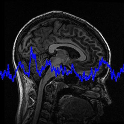
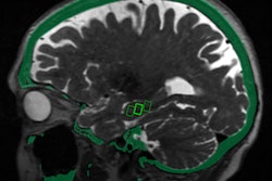
A treatment that uses ultrasound may be effective in relieving symptoms of essential tremor by targeting the affected area of the brain, according to a study published online November 20 in Neurology. The treatment, called focused ultrasound thalamotomy, essentially destroys the area of the brain causing the tremor.
More than 7 million people in the U.S. have essential tremor, according to the American Academy of Neurology. Currently, the most common treatment for the disorder when patients don't respond to medication is deep brain stimulation, a procedure that involves incisions and insertion of electrodes or probes into the patient's brain. Ultrasound thalamotomy would offer a less invasive treatment option and remains effective for approximately three years. It does, however, cause an irreversible brain lesion.
For the study, 56 patients received ultrasound thalamotomy and 20 received a fake treatment. The research team measured hand tremors, level of disability, and quality of life at the commencement of the study and after six months, one year, two years, and three years. By the three-year mark, the researchers noted a 50% improvement in hand tremors, a 56% improvement in disability, and a 42% improvement in quality of life.
While senior author Dr. Casey Halpern of Stanford University did note some limitations of the study, he concluded that "this treatment should be considered as a safe and effective option" for those living with essential tremor.






![Examples of ultrasound findings and techniques. (A) Images in a 39-year-old male patient with a mass in the left thigh. The mass is heterogeneous on the B-mode US image (compared with the patient in D) and showed increased microvascularity (superb microvascular imaging [SMI]) and shear-wave elastography (SWE) values. Undifferentiated pleomorphic sarcoma was diagnosed at biopsy (with pleomorphic rhabdomyosarcoma in surgical specimen). (B) Images in an 18-year-old male patient with a mass in the left leg. The mass is hypoechoic on the B-mode image, with no other findings suggestive of malignancy. The lesion is in contact with the cortex of the tibia, which is slightly irregular. CT revealed a doubtful anteromedial tibial erosion. The microvascular study demonstrated high vascularization, suggestive of malignancy. Periosteal Ewing sarcoma was diagnosed with both histologic and immunohistochemical confirmation. (C) Images in a 69-year-old female patient with a lump growing on the outside of the left leg. Multiple SWE examinations were performed (please note the high values obtained in the measurements, whereas the color map highlights the stiffness relative to adjacent tissues). SMI showed areas of increased vascularization to target for sampling. Undifferentiated spindle cell sarcoma was diagnosed at biopsy, with residual leiomyosarcoma in the surgical specimen after neoadjuvant therapy. (D) Images in a 56-year-old female patient with a mass in the right thigh. The mass is heterogeneous at both B-mode ultrasound (similar to patient A) and MRI (coronal T2-weighted spectral attenuated inversion recovery [SPAIR]; T1-weighted pre-contrast and postcontrast imaging), which even shows uptake after the administration of paramagnetic contrast material, which is traditionally suggestive of malignancy. Low values at SMI and elastography are suggestive of benignity. Spindle cell lipoma was diagnosed at biopsy, with atypical spindle cell lipomatous tumor in the surgical specimen.](https://img.auntminnie.com/mindful/smg/workspaces/default/uploads/2026/08/images-radiol250278fig2.APCFLSvX6p.jpg?auto=format%2Ccompress&fit=crop&h=112&q=70&w=112)


