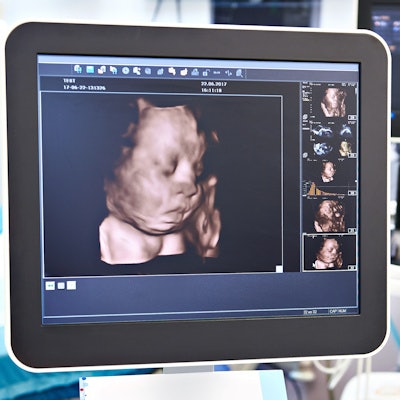
The woman who operated a keepsake ultrasound center in Louisiana that has been in the headlines this week is firing back at her critics, claiming she has done nothing wrong and that the negative media coverage is the result of a campaign orchestrated by a competitor.
Riley Dunaway and her Bump for Joy center were the targets of a critical news story on November 13 by television station WBRZ in Baton Rouge, LA. The story quoted a woman named Taylor Olivier, an expecting mother who visited Bump for Joy to get an ultrasound image of her unborn baby.
Olivier claims that Dunaway couldn't get a clear image of the fetus and asked her to perform jumping jacks to move it into a more suitable position. Olivier also claimed that Dunaway told her the wrong gender of the baby. When Olivier posted a complaint on a Facebook group on November 11, the post "went viral" and resulted in the WBRZ story, in which she claimed that 50 to 75 women had contacted her with concerns about their scans at Bump for Joy.
But in an email to AuntMinnie.com, Dunaway offered a rebuttal to several aspects of Olivier's story. She claims that Olivier originally left a "raving" review on Facebook and that jumping jacks are a standard method for repositioning a fetus when it is under 16 weeks gestation. Dunaway also claimed that the ultrasound image used in the WBRZ piece did not come from her center.
Dunaway said that she had received "raving reviews" in the six months that she had run Bump for Joy, during which she estimated that she scanned over 1,000 expecting women. She charged that a competing keepsake ultrasound center paid Olivier to go to her clinic and that the WBRZ reporter "has a long history of friendship with my competition."
"There is a legal battle going on right now and there will be light shed on the actual truth," Dunaway concluded.









![Examples of ultrasound findings and techniques. (A) Images in a 39-year-old male patient with a mass in the left thigh. The mass is heterogeneous on the B-mode US image (compared with the patient in D) and showed increased microvascularity (superb microvascular imaging [SMI]) and shear-wave elastography (SWE) values. Undifferentiated pleomorphic sarcoma was diagnosed at biopsy (with pleomorphic rhabdomyosarcoma in surgical specimen). (B) Images in an 18-year-old male patient with a mass in the left leg. The mass is hypoechoic on the B-mode image, with no other findings suggestive of malignancy. The lesion is in contact with the cortex of the tibia, which is slightly irregular. CT revealed a doubtful anteromedial tibial erosion. The microvascular study demonstrated high vascularization, suggestive of malignancy. Periosteal Ewing sarcoma was diagnosed with both histologic and immunohistochemical confirmation. (C) Images in a 69-year-old female patient with a lump growing on the outside of the left leg. Multiple SWE examinations were performed (please note the high values obtained in the measurements, whereas the color map highlights the stiffness relative to adjacent tissues). SMI showed areas of increased vascularization to target for sampling. Undifferentiated spindle cell sarcoma was diagnosed at biopsy, with residual leiomyosarcoma in the surgical specimen after neoadjuvant therapy. (D) Images in a 56-year-old female patient with a mass in the right thigh. The mass is heterogeneous at both B-mode ultrasound (similar to patient A) and MRI (coronal T2-weighted spectral attenuated inversion recovery [SPAIR]; T1-weighted pre-contrast and postcontrast imaging), which even shows uptake after the administration of paramagnetic contrast material, which is traditionally suggestive of malignancy. Low values at SMI and elastography are suggestive of benignity. Spindle cell lipoma was diagnosed at biopsy, with atypical spindle cell lipomatous tumor in the surgical specimen.](https://img.auntminnie.com/mindful/smg/workspaces/default/uploads/2026/08/images-radiol250278fig2.APCFLSvX6p.jpg?auto=format%2Ccompress&fit=crop&h=112&q=70&w=112)


