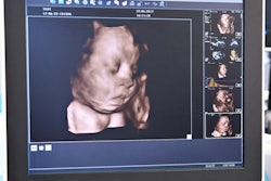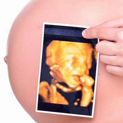
A group of expecting moms in Louisiana have discovered a hard lesson about the perils of keepsake ultrasound centers. The center apparently told dozens of women the wrong gender for their babies, and it even told one woman to do jumping jacks to enable a better view on ultrasound scans, according to a news report by television station WBRZ.
Bump for Joy opened in June 2019 in Prairieville, LA, as a "gender reveal, maternity, and children's boutique that also offers spray tanning." The center offered 2D, 3D, and 4D ultrasound scans to pregnant women, and it pointed out that it could determine the gender of babies at 13 weeks gestation.
Bump for Joy also offered a "Baby Blues Package" for expecting mothers who had experienced loss, had a miscarriage, or had infertility issues. The package included an ultrasound scan once a week for 15 minutes, according to news reports of the center's opening. Scans were performed by owner Riley Dunaway, who told reporters she had "completed a course" and had two years of supervised training in ultrasound.
But mothers-to-be soon began reporting complaints about the keepsake ultrasound center. One woman who was profiled by WBRZ said that she paid Dunaway $70 for scans prior to a gender-reveal party and was told she was having a boy.
She described her experience at Bump for Joy as "awkward," as Dunaway apparently had difficulty getting a good image of the fetus. Also, Dunaway told the expecting mom halfway through the 30-minute session to get up and do jumping jacks to "allow for a better view of the baby," WBRZ reported.
After reading complaints about Bump for Joy on a Facebook page, the woman got a second opinion at 16 weeks -- and scans revealed she was pregnant with a girl.
What's more, the woman said she was contacted by 50 to 75 women who said that Bump for Joy had given them incorrect information on the gender of their baby or medical advice that turned out to be incorrect.
When a reporter from WBRZ visited Bump for Joy's offices, she found the lights off and no one answered the door. Reached by phone, Dunaway told WBRZ that she decided to close the business after the negative publicity.