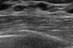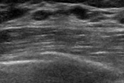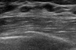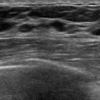
The automated breast volume scanner (ABVS) performs just as well as handheld ultrasound for distinguishing between malignant and benign breast lesions, according to a literature review published in the August issue of Ultrasound in Medicine and Biology.
The findings suggest that clinicians can trust ABVS as an alternative to handheld ultrasound -- which can be affected by user skill variability -- and that it shows promise as a breast imaging technique for both screening and diagnosis, wrote co-authors Dr. Liang Wang and Zhen-Hong Qi of Peking Union Medical College Hospital in Beijing.
"Conventional handheld ultrasound has several inherent limitations, including the operator dependence and the time required for whole-breast examination," they wrote (Ultrasound Med Biol, August 2019, Vol. 45:8, pp. 1874-1881). "In contrast, the automated breast volume scanner has several advantages over handheld ultrasound, such as higher reproducibility, less operator dependence, and less time required for image acquisition."
Most radiologists continue to use handheld ultrasound to characterize breast lesions identified on mammography or other screening modalities, and the diagnostic performance of ABVS is controversial, Wang and Qi noted. To better assess this, the two conducted a literature review of PubMed, Embase, and Cochrane Library databases.
Their research included nine studies with 1,985 lesions from 1,774 patients; of the lesions, 628 were malignant and 1,357 were benign. Wang and Qi evaluated the following performance measures for the two techniques: sensitivity, specificity, positive and negative likelihood ratios, and diagnostic odds ratios.
Across all measures, ABVS performed comparably to handheld ultrasound for distinguishing benign from malignant breast lesions, the researchers found.
| Comparison of handheld ultrasound and ABVS for distinguishing benign from malignant breast lesions | ||
| Performance measure | Handheld ultrasound | ABVS |
| Sensitivity | 90.6% | 90.8% |
| Specificity | 81% | 82.2% |
| Positive likelihood ratio | 5.22 | 5.39 |
| Negative likelihood ratio | 0.11 | 0.10 |
| Diagnostic odds ratio | 52.60 | 61.68 |
| Area under the receiver operating characteristic curve | 0.94 | 0.93 |
"On the basis of our data, the ABVS and handheld ultrasound exhibited similar sensitivity and specificity in the differentiation of malignant and benign breast lesions," Wang and Qi wrote.
They cautioned that these results were based on grayscale ultrasound interpretation, meaning that only morphological features were displayed, rather than functional information that elastography and Doppler ultrasound offer.
"Both elastography and Doppler ultrasound can provide independent diagnostic information in addition to the grayscale imaging, which could significantly improve the diagnostic accuracy for breast ultrasound," they wrote.
But ABVS does have an advantage over handheld ultrasound that could make it a valuable tool in clinical practice: its ability to gather additional morphological information on the reconstructed coronal plane.
"The retraction phenomenon on the coronal plane of the ABVS is regarded as a highly diagnostic feature in the differentiation of benign and malignant breast lesions," the researchers wrote. "Therefore, it seems reasonable that the ABVS may be superior to handheld ultrasound in differential diagnosis with the help of coronal reconstruction."