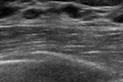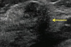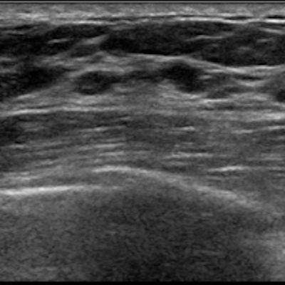
Using a novel ultrasound technique called ultrasensitive ultrasound microvessel imaging (UMI) with conventional ultrasound significantly improves the diagnostic accuracy of BI-RADS classification of breast lesions, according to a study to be published in the December issue of Ultrasound in Medicine and Biology.
And improved BI-RADS categorization could translate into fewer unnecessary breast biopsies, wrote a team led by Ping Gong, PhD, of the Mayo Clinic in Rochester, MN.
"The microvessel information provided by ultrasound microvessel imaging may add clinical value to supplemental ultrasound screening adjunct to mammography in reducing unnecessary and missed biopsies," the researchers wrote (Ultrasound Med Biol, December 2019, Vol. 45:12, pp. 3128-3136).
Reducing false positives
Combining ultrasound with mammography improves diagnostic sensitivity in women with dense tissue compared with mammography alone, Gong and colleagues noted. But the combination can also increase false positives due to the low specificity of ultrasound -- leading to more unnecessary biopsies. That's why improving ultrasound's performance is important.
"In contrast to normal tissue and benign tumors, malignant tumors typically present a vessel pattern with chaotic distribution, irregular branches, and penetrating peripheral vasculature," the group wrote. But the "clinical reliability of conventional Doppler is undermined by limited vessel detection sensitivity. An ultrasensitive ultrasound microvessel imaging technique ... [provides] advanced vessel sensitivity ... without using ultrasound contrast agents."
Instead of the line-by-line scanning of conventional Doppler ultrasound, UMI is based on "ultrafast plane wave imaging ... that leads to at least 10 times more ultrasound frames for blood flow detection than conventional Doppler," the group wrote. This rapid imaging allows for a much higher sensitivity for visualizing microvessels, which helps distinguish between malignant and benign lesions, study co-author Shigao Chen, PhD, also of the Mayo Clinic, told AuntMinnie.com.
"Conventional ultrasound has low sensitivity for detecting very small vessels, and thus may miss the microvessels in the tumor which provide important information for diagnosis," he said. "This technology uses a newer ultrasound scanner with very high imaging frame rates so that more data can be acquired to boost sensitivity, and it uses more advanced signal processing to better remove tissue signal -- which is orders of magnitudes higher than blood signal -- to reveal the underlying microvessels," he said.
The researchers sought to explore the feasibility of UMI for assessing breast tumor microvessel distribution and to compare the performance of UMI and conventional ultrasound in differentiating between benign and malignant lesions. The study included 44 women with 51 breast masses, all of whom were imaged with conventional ultrasound and the combination of conventional ultrasound and UMI. Of the 51 masses, 28 were malignant and 23 were benign.
Gong's group found that adding UMI to conventional ultrasound significantly improved the visualization of small vessels compared with Doppler ultrasound alone across a variety of performance measures and that the microvessel structures UMI depicted were associated with benign or malignant tumor characteristics.
| Performance of conventional ultrasound alone compared with ultrasound plus UMI | ||
| Performance measure | Conventional ultrasound | Ultrasound plus UMI |
| Sensitivity | 96.4% | 100% |
| Specificity | 17.4% | 47.8% |
| Positive predictive value | 58.7% | 70% |
| Negative predictive value | 80% | 100% |
| Diagnostic accuracy | 60.8% | 76.5% |
The study also found that the diagnostic accuracy of correct BI-RADS categories improved by 16 percentage points with the combination of conventional ultrasound and UMI compared with ultrasound alone.
"This improvement indicates the potential of UMI in reducing unnecessary benign biopsies and avoiding missed malignant biopsies," Gong and colleagues wrote.
Keep the research coming
More research using this ultrasound technique is needed, Chen told AuntMinnie.com.
"If this preliminary research is confirmed by larger studies, it will imply that information about the microvasculature of the tumor obtained by ultrasensitive Doppler ultrasound can be used to differentiate benign from malignant tumors and thus reduce unnecessary biopsy," he said. "In addition, the technology may be useful for early evaluation of treatment effects to guide adjustment of chemotherapy or medical therapy regimens and avoid unnecessary side effects of ineffective therapies."