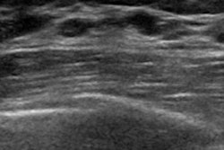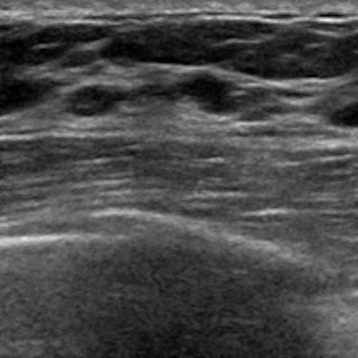
Both automated breast ultrasound (ABUS) and traditional handheld ultrasound can significantly improve breast cancer detection when used as an adjunct to mammography in women with dense breasts. But ABUS yields better diagnostic performance, according to research presented at RSNA 2019 in Chicago.
In a study involving over 1,200 women, a team of researchers led by Dr. Mengmeng Jia of the Chinese Academy of Medical Sciences in Beijing found that ABUS produced a higher specificity, positive predictive value, and area under the curve (AUC) than handheld ultrasound.
Breast cancer is the most commonly diagnosed cancer in Chinese women, but less than 1% of cases are detected by screening, according to Jia. Compared with Western countries, Chinese women also have a higher proportion of dense breasts, for which mammography is less sensitive, she said. As a result, these patients need adjunctive imaging modalities such as ultrasound, digital breast tomosynthesis, or MRI.
Traditional handheld ultrasound is inexpensive, safe, and suitable for dense breasts. But it's also labor intensive and highly dependent on the operator. ABUS, on the other hand, is specifically designed for finding cancer in dense breast tissue. It's also reproducible due to its operator-independent acquisition method, Jia noted.
"Most importantly, the image acquisition can be separated from interpretation," she said. "That means that images can be taken by the operator and then interpreted by radiologists [at another location]. That is helpful in resource-limited areas [where there are not enough qualified radiologists]."
The researchers sought to evaluate the diagnostic performance of ABUS and handheld ultrasound as an adjunct to mammography in women ages 40 to 69. They also wanted to assess the performance of both methods in mammography-negative dense breasts.
The team enrolled 1,266 women ages 40 to 69 in a multicenter study involving five tertiary hospitals. All women received mammography, as well as handheld ultrasound and ABUS. Of the 1,266 women, 958 were deemed to have dense breasts.
Overall, sensitivity increased from 87.3% with mammography alone to 96.9% for both handheld ultrasound and ABUS. Negative predictive value also increased from 95.5% to 98.8% with handheld ultrasound and 98.8% for ABUS. Mammography alone had an AUC of 0.88, compared with an AUC of 0.92 for mammography and handheld ultrasound and 0.93 for mammography and ABUS.
In women with mammographically negative dense breasts, ABUS and handheld ultrasound detected 31 additional cases of breast cancer. The techniques had comparable sensitivity and negative predictive value, but ABUS had a higher specificity, positive predictive value, and AUC, according to the researchers.
| Performance in women with mammographically negative dense breasts | ||
| Handheld ultrasound | ABUS | |
| Sensitivity | 31/33 (93.9%) | 31/33 (93.9%) |
| Specificity | 619/665 (93.1%) | 635/665 (95.5%) |
| Positive predictive value | 31/77 (40.3%) | 31/61 (50.8%) |
| Negative predictive value | 619/621 (98.8%) | 635/637 (99.7%) |
| Area under the curve | 0.935 | 0.947 |
"More studies are [now] needed to [further] evaluate the performance of adjunctive ultrasonography, including ABUS and handheld ultrasound, in [resource]-limited areas," Jia concluded.