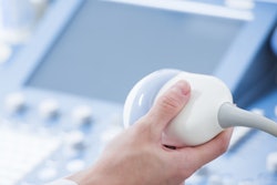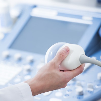
Healthcare personnel in Malawi used ultrasound to diagnose tuberculosis (TB) in patients infected with HIV with the help of a teleradiology link that sent images to a U.S.-based radiologist, according to a report published online September 26 in the Journal of the American College of Radiology.
The findings are good news for physicians and other providers serving in underresourced healthcare environments and in developing countries, wrote the team led by Dr. Kristin Schwab from the University of California, Los Angeles (UCLA).
"The diagnosis of tuberculosis in patients infected with HIV can be challenging in resource-limited settings," the group wrote. "Extrapulmonary TB is particularly underdiagnosed, with data suggesting that half of patients with disseminated TB are not diagnosed until autopsy. Ultrasound represents a promising diagnostic modality because of its low cost, widespread availability, and minimal risks to the patient."
Schwab's group conducted a study in Malawi that sought to investigate whether training medical staff on a protocol called "focused assessment with sonography for HIV-associated TB" (FASH) and providing feedback and support via a remotely based U.S. radiologist would prove valuable for diagnosing the disease in this population.
Eleven physicians, radiographers, and medical assistants in Malawi participated in four-day training courses taught by two UCLA physicians. The courses were held at three sites: a medical center, a district hospital, and a mission hospital; each session included four or five participants and three groups were trained weekly. Of these 11 trainees, nine were physicians, one was a radiographer, and one was a medical assistant.
After being trained, staff performed ultrasound assessments for HIV-associated tuberculosis on control subjects who were HIV-positive but asymptomatic for TB (enrolled between February and March 2016), as well as a set of "case" subjects who were HIV-positive and had signs of TB (enrolled between March 2016 and April 2017). Of the subjects, 75 were in the control group and 108 were in the case group.
The trainees in the field acquired nine ultrasound images from each patient, interpreted the images, and sent them to a remote U.S. radiologist with ultrasonography expertise. Schwab's team then assessed concordance between the interpretations of the trainees and those of the expert radiologists.
After the training and teleradiology support, the personnel in Malawi identified 92% of abnormalities also identified by the expert radiologist in the U.S.
"Our ... study suggests that both physician and nonphysician clinicians can learn the technical skills of ultrasound image acquisition after a short four-day training course followed by focused teleultrasonography feedback," the authors concluded.
"[The findings have] the potential not only to improve the diagnosis of extrapulmonary TB in the global health setting, but also to inspire the creation of future protocols using teleultrasonography to harness radiology expertise for training in resource-limited settings," they wrote.