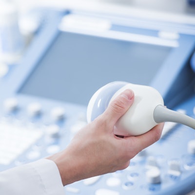
Ultrasound can help clinicians predict whether arteriovenous fistulas (AVFs) -- surgically created sites that connect a patient's vein and artery -- will prove effective for accessing the bloodstream for dialysis, according to a study published online October 11 in the Journal of the American Society of Nephrology.
Creating an arteriovenous fistula is the preferred method for gaining entry into the bloodstream for hemodialysis. But up to 60% of fistulas fail to mature properly and are unable to be used for maintenance hemodialysis, wrote a team led by Dr. Michelle Robbin from the University of Alabama at Birmingham. For it to work, the AVF's diameter and blood flow must increase after it is created.
"An easily palpable AVF with a shallow depth from the skin can be cannulated more readily than a deeper one," the group wrote.
Clinicians have long used physical examination to determine whether an AVF has successfully matured, and this works in up to 80% of patients. But physical exams are subject to variation in staff skill levels. What's really needed is an accurate, noninvasive, and inexpensive test that can evaluate whether an arteriovenous fistula has progressed toward clinical maturation, according to Robbin and colleagues.
That's where ultrasound comes in.
"We investigated the relationships of AVF blood flow, diameter, and depth, measured postoperatively ... [to determine] whether these ultrasound measurements could predict clinical maturation accurately enough for practical use," the group wrote.
The study included 602 patients with chronic kidney disease who had surgery to create a single upper-extremity AVF between March 2010 and August 2013. The patients underwent postsurgical ultrasound at one day, two weeks, and six weeks. Robbin and colleagues used odds ratios to calculate the effectiveness of the ultrasound evaluations for predicting AVF maturation; success was defined as being able to use the AVF for 75% of dialysis sessions over a continuous four-week period.
The investigators found that the three ultrasound assessments were effective in predicting the clinical maturation of the AVFs across a variety of patient characteristics, including age, sex, race, AVF location, dialysis status, diabetes status, and body mass index.
| Association of AVF maturation with ultrasound assessments | ||
| Ultrasound assessment | Odds ratio | p-value |
| AVF blood flow | ||
| Week 1 | 5.88 | < 0.001 |
| Week 2 | 4.24 | < 0.001 |
| Week 6 | 8.81 | < 0.001 |
| Vein diameter | ||
| Week 1 | 3.56 | < 0.001 |
| Week 2 | 5.53 | < 0.001 |
| Week 6 | 4.74 | < 0.001 |
| Vein depth | ||
| Week 1 | 0.56 | 0.06 |
| Week 2 | 0.37 | 0.004 |
| Week 6 | 0.38 | 0.006 |
| Arterial diameter | ||
| Week 1 | 1.64 | 0.21 |
| Week 2 | 2.66 | 0.005 |
| Week 6 | 3.21 | 0.002 |
The study suggests that ultrasound can offer patients and their clinicians a noninvasive way to assess whether an AVF will be as effective as it can be, Robbin and colleagues wrote.
"We found that these three ultrasound measurements fully accounted for the associations of both unassisted and overall clinical maturation with the case-mix variables," the researchers concluded.