Ultrasound scans weren't initially able to detect microcephaly in the fetus of a pregnant woman infected with the Zika virus, according to a case report published on March 30 in the New England Journal of Medicine. Signs of Zika's most feared fetal defect only became evident more than a month after infection.
A multi-institutional team led by Dr. Rita Driggers of Johns Hopkins University Hospital shared the case of a 33-year-old Finnish woman in her 11th week of gestation who had acquired the Zika virus while vacationing with her husband in Mexico, Guatemala, and Belize in late November 2015.
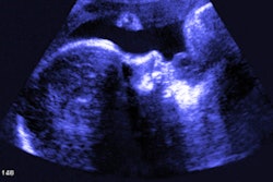
Fetal ultrasound performed at 13, 16, and 17 weeks of gestation (one, four, and five weeks, respectively, after Zika symptoms were resolved) found no evidence of the microcephaly or intracranial calcifications that are associated with the virus. However, ultrasound showed a decrease in fetal head circumference from the 47th percentile at 16 weeks to the 24th percentile at 20 weeks.
"Negative ultrasonographic studies during this [latency] period would be falsely reassuring and might delay critical time-sensitive decision-making," the authors wrote."Serial ultrasonographic measurements of head circumference may provide useful predictive information. The superior soft-tissue resolution of fetal brain MRI might be more sensitive to developmental and encephaloclastic changes, thereby expediting the detection of evolving fetal brain anomalies."
Thorough assessment
Healthcare providers detected the virus in the woman at 16 weeks of gestation using a blood assay, which identified Central American epidemic strains of the Zika virus. Based on those results, the patient sought a more thorough fetal assessment, according to the researchers.
An ultrasound scan detected abnormal intracranial anatomy in the fetus at 19 weeks of gestation, including a thin appearance of the cerebral mantle with increased extra-axial spaces, as well as an enlargement of both frontal horns that raised concern about intraventricular hemorrhage, according to the team.
The ultrasound also showed dilation and upward displacement of the third ventricle, dilatation of the frontal horns of the lateral ventricles, concave medial borders of the lateral ventricles, and the absence of the cavum septum pellucidum suggesting agenesis of the corpus callosum. Gestational age was measured at the 24th percentile, although the rest of the fetal anatomy was normal.
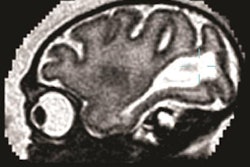
The patient also received an MRI scan at 20 weeks of gestation, which demonstrated diffuse atrophy of the cerebral mantle. The atrophy was most severe in the frontal and parietal lobes, according to the group. The researchers noted that the cerebral mantle's normal lamination pattern was absent and the subplate zone was lately undetectable.
What's more, the corpus callosum had an anterior-posterior length of 14 mm, which was significantly shorter than the expected range of 18 mm to 22 mm for the gestational age. The MRI also showed that the cavum septum pellucidum was very small and that the lateral ventricles had a mildly enlarged transverse diameter of 2.5 mm -- the average measurement for the lateral ventricle is 1.75 mm for this gestational age, with a range of 1.1 mm to 2.3 mm. In addition, the choroid plexus had an unusually prominent volume, although without evidence of hemorrhage, the group reported.
Grave prognosis
Based on the grave prognosis, the patient decided to terminate the pregnancy at 21 weeks of gestation, according to the researchers. Postmortem analysis showed that the highest Zika viral loads were in the fetal brain, with substantial viral loads in the placenta, fetal membranes, and umbilical cord. The group also found lower amounts of Zika virus RNA in the fetal muscle, liver, lung, and spleen; amniotic fluid at the time of termination was positive for Zika virus RNA with low viral counts.
The study highlights the possible importance of RNA serum testing for the Zika virus in pregnant women beyond the first week after the onset of symptoms, according to the researchers. In addition, imaging techniques such as serial fetal ultrasonography and fetal brain MRI could be used in suspected cases. Future studies should provide additional evidence of markers signifying Zika infection, they said.