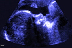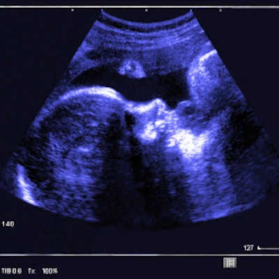
Performing ultrasound scans late in pregnancy helps women avoid undiagnosed breech presentation of their babies, translating to improved clinical outcomes, lower rates of emergency cesarean sections (C-sections), and perhaps even lower healthcare costs, according to a study published April 16 in PLOS Medicine.
The findings are good news not only for women and their babies but also for the healthcare system, wrote a team led by David Wästlund of the University of Cambridge in the U.K.
"According to our estimates, universal late pregnancy ultrasound in nulliparous women would virtually eliminate undiagnosed breech presentation, would be expected to reduce fetal mortality in breech presentation, and would be cost-effective if fetal presentation could be assessed for less than 19.80 pounds [$25.95 U.S.] per woman," the group wrote.
Fetal breech presentation increases the risk of complications for the baby and the mother, Wästlund and colleagues noted. Typically, a baby's position is assessed by palpating the woman's abdomen, but this technique's sensitivity varies by practitioner.
"Despite the relative ease with which breech presentation can be identified through ultrasound screening, the assessment of fetal presentation at term is often based on clinical examination only," the researchers wrote. "Due to limitations in this approach, many women present in labor with an undiagnosed breech presentation."
The investigators performed screening ultrasound at 36 weeks gestation in 3,879 English women having first pregnancies between January 2008 and July 2012. Of these, 179 (4.6%) were diagnosed with breech presentation. In more than half of those (54%), breech presentation had not been suspected prior to labor.
Women with babies in the breech position were offered a procedure called an external cephalic version (ECV) to try to turn the baby; for those who did not want this procedure or for whom it did not work, a C-section was scheduled.
The investigators also estimated the cost of universal late pregnancy ultrasound scans using data from the English National Health Service (NHS) to compare birth outcomes of breech pregnancies screened with and without ultrasound.
The ECV procedure was attempted in 84 (46.9%) of the women with breech babies and was successful in 12 (14.3%). Of the 179 women with breech babies, the researchers found the following:
- 10.6% delivered vaginally (following either a planned or spontaneous version).
- 61.5% delivered via elective C-section.
- 27.9% delivered via emergency C-section (due to labor starting before the scheduled cesarean date).
"No woman in the cohort had a vaginal breech delivery or experienced an intrapartum cesarean for undiagnosed breech," the researchers noted.
Wästlund and colleagues estimated that routine late pregnancy ultrasound could prevent nearly 15,000 undiagnosed breech presentations, more than 4,000 emergency C-sections, and seven to eight baby deaths per year. But the effect of the intervention on healthcare costs needs more research, the group wrote: "If universal ultrasound could be provided for less than 12.90 pounds [$16.91] per scan, the policy would also be cost saving." But it's unclear if this is possible.
"If this procedure could be implemented into routine care, for example, by midwives conducting a routine [scan at 36 weeks gestation] and using a portable ultrasound system, it is likely to be cost-effective," the group concluded. "Such a program would be expected to reduce the consequences to the child of undiagnosed breech presentation, including morbidity and mortality."