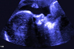
How often does a repeat screening fetal ultrasound study performed because of an incomplete initial exam actually detect abnormal fetal anatomy? Not very often, according to research published in the December issue of the Journal of Ultrasound in Medicine.
In a retrospective study involving more than 16,000 fetal anatomic screening ultrasound exams, researchers from Yale University found that nearly one in eight of these exams was deemed incomplete. Among the resulting repeat exams performed, only 0.5% led to the detection of abnormal fetal anatomy that hadn't been seen before, according to lead author Dr. Mark Silvestri and colleagues.
 Dr. Mark Silvestri from Yale University.
"Having women come back to repeat the exam may not be universally beneficial, and expecting mothers should understand the likely outcomes of a repeat exam," Silvestri told AuntMinnie.com.
Clinical utility?
The Yale research project stemmed from an experience Silvestri had as an ob/gyn resident when he and his wife were expecting their first child.
"At our anatomy scan, we were told they couldn't quite see the profile and nose adequately, and they wanted us to come back for a repeat ultrasound," he said. "But if I showed you one of the pictures we took home that day, it was a pretty great picture of my son's profile and nose -- even if it wasn't technically adequate by the exam standards."
Although they wound up not going back for the repeat ultrasound, Silvestri was interested in learning how often this situation occurs for other parents and what the outcomes are when women do return for the repeat studies.
"Healthcare resources are finite," Silvestri said. "There are many health outcomes -- including obstetric outcomes -- in which our country is underperforming, so it is important not to spend our limited resources on unnecessary health services."
The researchers identified all initial fetal anatomic ultrasound exams performed at the Yale-New Haven Hospital obstetric ultrasound center between January 1, 2009, and December 31, 2013. Of the 16,300 exams included in the study, 14,143 (86.9%) had complete anatomic visualization, of which 877 (6.2%) were abnormal.
The remaining 2,157 (13.2%) were deemed to have incomplete visualization of fetal anatomy (J Ultrasound Med, December 2016, Vol. 35:12, pp. 2665-2673).
Repeat recommendations
Of these 2,157 exams, 302 (14%) received a recommendation for targeted repeat imaging because of a suspected or confirmed abnormality on the initial imaging along with the incomplete visualization. In addition, 17 cases (0.8%) involved a situation such as fetal demise for which the patient was not eligible for repeat imaging.
Of the remaining 1,838 women with an otherwise normal examination, 1,681 (91.5%) were recommended for repeat ultrasound; 1,560 (92.8%) of these women subsequently received the repeat exam.
"In our study, one in eight pregnant women had an anatomy ultrasound exam that was incomplete (it did not completely visualize all necessary body parts of the baby)," Silvestri said. "Nearly all of these women were asked to come back for another ultrasound exam, and almost all of them did. If these numbers were consistent across the U.S., that would equate to approximately 500,000 pregnant women each year who are asked to return for a repeat ultrasound exam for this reason."
Of the 1,560 repeat ultrasound exams, 1,380 (88.5%) were completed and had normal findings for the previously incompletely visualized anatomical components. Notably, an additional 172 (11%) still had incomplete visualization of the same structures. Only eight (0.5%) of the repeat exams showed a suspected or confirmed abnormality that had previously been incompletely visualized, according to the group.
"Based on our findings, nine times out of 10 the fetal body parts will be found to be normal, one time out of 10 they will remain poorly visualized, and one time out of 200 they will be found to be abnormal," Silvestri said.
There were six major abnormalities: two cases of multicystic dysplastic kidney, one case of tetralogy of Fallot, one case of aortic stenosis, one case of hypoplastic left heart, and one case of cerebral ventriculomegaly. Two minor abnormalities were found: one case of postaxial polydactyly and one case of choroid plexus cyst.
Silvestri noted that all ultrasounds were performed at a single obstetric ultrasound center. As a result, their findings might not apply universally to all other settings.
A more judicious approach
The research shows that these repeat exams may not always be needed, Silvestri said.
"I would propose that a more thoughtful and judicious approach should be used in recommending them," he said. "It is exciting to learn that our excess caution in recommending these repeat studies might not be necessary, and that we could instead tailor our recommendations to a patient's particular clinical situation."
Given the litigious environment and fee-for-service reimbursement that pays for each additional ultrasound exam, Silvestri acknowledged that it may be challenging to persuade physicians to forego recommending repeat ultrasounds.
"Ideally there would be a shift in some of the structural reasons that have likely contributed to these repeat exams becoming common," Silvestri said. "Specialty societies could help, however, by providing guidelines that advocate a more judicious approach to repeating anatomy ultrasounds."