(Radiology Review) Several previously undescribed sonographic signs indicating ruptured abdominal aortic aneurysm (AAA) were recently identified by Italian radiologists. Traditionally, sonography has been used to confirm AAA, and CT used to confirm rupture because of its high accuracy in demonstrating retroperitoneal hemorrhage in patients with suspected rupture. This latest retrospective study indicates that sonography could demonstrate direct and indirect signs of aneurysm rupture and expedite surgery in unstable patients, thus avoiding a medical catastrophe.
In the August issue of the Journal of Ultrasound in Medicine, Drs. Orlando Catalano and Alfredo Siani from the Istituto Pascale in Naples outlined the ultrasound findings seen in patients with ruptured AAA, in particular three new findings that can be used to diagnose ruptured AAA.
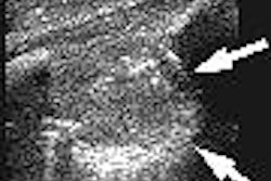
Surgical or CT demonstration of aneurysm rupture was found in 29 of 388 consecutive patients with suspected AAA. A transverse aortic diameter of greater than 3 cm was used to diagnose AAA. "Findings recognized among 29 positive cases included AAA deformation, luminal thrombus inhomogeneity, clear interruption of a luminal thrombus, retroperitoneal hematoma, and hemoperitoneum," the authors reported. Additionally, three new signs included a layer of intraluminal floating thrombus layer, a parietal hypoechoic focus due to discontinuity of the aneurysm wall, and a para-aortic hypoechoic area adjacent to the interrupted wall.
"Aside from AAA deformation and thrombus heterogeneity, no other signs were recognized among subjects with a nonruptured aneurysm," they reported. Peripheral intraluminal thrombus was seen in 298 patients, and this usually was eccentric (85%) in location.
The authors described the three newly reported sonographic signs in great detail, and cautioned that the floating thrombus layer should not be confused with aortic dissection, which is characterized by an extended echoic intimal flap, attached on both ends to the aortic wall. "Instead, we describe the floating layer as an irregular flap, detached focally from the aneurysm thrombus on one side and freely floating within the aneurysm lumen."
They described an AAA outer wall tear as "a focal hypoechoic area on the side of retroperitoneal hemorrhage, with interruption of the aneurysm's calcified rim." The para-aortic hypoechoic area, demonstrated a "small anechoic focus immediately adjacent to the AAA wall, surrounded by retroperitoneal hematoma and indicating the location of the freshest area of bleeding."
"In the proper clinical setting, sonographic detection of an AAA should prompt further imaging (CT examination) or immediate surgery. However, we also think that current state-of-the-art sonographic equipment allows recognition of many signs of rupture itself," the authors concluded.
"Ruptured Abdominal Aortic Aneurysm: Categorization of Sonographic Findings and Report of 3 New Signs"
Orlando Catalano, MD, and Alfredo Siani, MD
Department of Radiology, Istituto Pascale
Via Crispi 92
I-80121 Naples, Italy
J Ultrasound Med 2005 (August); 24:1077-1083
By Radiology Review
August 15, 2005
Copyright © 2005 AuntMinnie.com
![Examples of ultrasound findings and techniques. (A) Images in a 39-year-old male patient with a mass in the left thigh. The mass is heterogeneous on the B-mode US image (compared with the patient in D) and showed increased microvascularity (superb microvascular imaging [SMI]) and shear-wave elastography (SWE) values. Undifferentiated pleomorphic sarcoma was diagnosed at biopsy (with pleomorphic rhabdomyosarcoma in surgical specimen). (B) Images in an 18-year-old male patient with a mass in the left leg. The mass is hypoechoic on the B-mode image, with no other findings suggestive of malignancy. The lesion is in contact with the cortex of the tibia, which is slightly irregular. CT revealed a doubtful anteromedial tibial erosion. The microvascular study demonstrated high vascularization, suggestive of malignancy. Periosteal Ewing sarcoma was diagnosed with both histologic and immunohistochemical confirmation. (C) Images in a 69-year-old female patient with a lump growing on the outside of the left leg. Multiple SWE examinations were performed (please note the high values obtained in the measurements, whereas the color map highlights the stiffness relative to adjacent tissues). SMI showed areas of increased vascularization to target for sampling. Undifferentiated spindle cell sarcoma was diagnosed at biopsy, with residual leiomyosarcoma in the surgical specimen after neoadjuvant therapy. (D) Images in a 56-year-old female patient with a mass in the right thigh. The mass is heterogeneous at both B-mode ultrasound (similar to patient A) and MRI (coronal T2-weighted spectral attenuated inversion recovery [SPAIR]; T1-weighted pre-contrast and postcontrast imaging), which even shows uptake after the administration of paramagnetic contrast material, which is traditionally suggestive of malignancy. Low values at SMI and elastography are suggestive of benignity. Spindle cell lipoma was diagnosed at biopsy, with atypical spindle cell lipomatous tumor in the surgical specimen.](https://img.auntminnie.com/mindful/smg/workspaces/default/uploads/2026/08/images-radiol250278fig2.APCFLSvX6p.jpg?auto=format%2Ccompress&fit=crop&h=100&q=70&w=100)


![Examples of ultrasound findings and techniques. (A) Images in a 39-year-old male patient with a mass in the left thigh. The mass is heterogeneous on the B-mode US image (compared with the patient in D) and showed increased microvascularity (superb microvascular imaging [SMI]) and shear-wave elastography (SWE) values. Undifferentiated pleomorphic sarcoma was diagnosed at biopsy (with pleomorphic rhabdomyosarcoma in surgical specimen). (B) Images in an 18-year-old male patient with a mass in the left leg. The mass is hypoechoic on the B-mode image, with no other findings suggestive of malignancy. The lesion is in contact with the cortex of the tibia, which is slightly irregular. CT revealed a doubtful anteromedial tibial erosion. The microvascular study demonstrated high vascularization, suggestive of malignancy. Periosteal Ewing sarcoma was diagnosed with both histologic and immunohistochemical confirmation. (C) Images in a 69-year-old female patient with a lump growing on the outside of the left leg. Multiple SWE examinations were performed (please note the high values obtained in the measurements, whereas the color map highlights the stiffness relative to adjacent tissues). SMI showed areas of increased vascularization to target for sampling. Undifferentiated spindle cell sarcoma was diagnosed at biopsy, with residual leiomyosarcoma in the surgical specimen after neoadjuvant therapy. (D) Images in a 56-year-old female patient with a mass in the right thigh. The mass is heterogeneous at both B-mode ultrasound (similar to patient A) and MRI (coronal T2-weighted spectral attenuated inversion recovery [SPAIR]; T1-weighted pre-contrast and postcontrast imaging), which even shows uptake after the administration of paramagnetic contrast material, which is traditionally suggestive of malignancy. Low values at SMI and elastography are suggestive of benignity. Spindle cell lipoma was diagnosed at biopsy, with atypical spindle cell lipomatous tumor in the surgical specimen.](https://img.auntminnie.com/mindful/smg/workspaces/default/uploads/2026/08/images-radiol250278fig2.APCFLSvX6p.jpg?auto=format%2Ccompress&fit=crop&h=112&q=70&w=112)