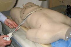
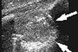
Sonography can play a role in detecting residual primary breast malignancy in patients receiving neoadjuvant chemotherapy, yielding high sensitivity for tumors 7 mm or larger, according to an article published in the July issue of the Journal of Ultrasound in Medicine.
"Radiologists and sonographers who perform breast sonography should be aware of the changing appearance of breast cancers during chemotherapy and of the advantages and limitations of sonography in assessing tumor response," wrote a research team from the University of Michigan in Ann Arbor, MI; Oakwood Healthcare System in Dearborn, MI; and Primary Children's Medical Center in Salt Lake City, UT.
The study team prospectively evaluated 34 women with early-stage operable breast cancers (mean maximum size of 2.4 cm) who received neoadjuvant chemotherapy with doxorubicin and docetaxel (JUM, July 2005, Vol. 24:7, pp. 885-895).
Targeted whole-volume sonography was performed using a Logiq 700 scanner (GE Healthcare, Chalfont St. Giles, U.K.) on the tumor sites both before and after chemotherapy to assess mass size, color pixel speed-weighted density, and American College of Radiology Breast Imaging Reporting and Data System (BI-RADS) sonographic characteristics.
Following chemotherapy, tumor sites were excised by lumpectomy or mastectomy.
Images were acquired at approximately 0.5-mm intervals in grayscale and color Doppler imaging modes.
Of the 34 patients, three (11.3%) had a complete histologic response. Following chemotherapy, correlation was r = 0.716 between final histologic and sonographic sizes.
Compared with histologic residual tumors, sonography produced four false-negative results, three false-positive results, and 27 true-positive results for sensitivity of 87%.
There were no false-negative results among a subgroup of tumors of 7 mm and larger. The three false-positive cases were histologic fibrosis or biopsy changes, according to the authors.
In other findings, the researchers found mean speed-weighted density to be 0.015 before chemotherapy and 0.0082 afterward (p = 0.03). After chemotherapy, vascularity was less common within (p = 0.06) or adjacent to (p = 0.009) masses or in tumor sites (p = 0.05).
The study team did not discover any grayscale or vascular characteristic that predicted tumor response, although statistical evaluation was limited by the small number of complete responders. Also, grayscale characteristics commonly change after chemotherapy, but not in a predictable way, the researchers said.
"Some changes may be due to shrinking tumor size, development of fibrosis or cellular necrosis, and the tumor's becoming multifocal," they wrote. "Therefore, the appearance of the tumor after chemotherapy may be rather different, and identifying the location with tissue markers is important for postchemotherapy localization. Grayscale characteristics also do not distinguish the false-positive results from the true-positive residual tumors."
After chemotherapy, maximum tumor size at histologic comparison correlated quite well with the final sonographic size among cancers with a maximal size of 7 mm or larger, the authors noted. Below that threshold size, sonography yielded false-negative results.
"On the basis of our findings and those of other studies, we hypothesize that the histologic effects of docetaxel may contribute to false-negative grayscale sonographic results, and that antiangiogenic effects make a decrease in vascularity a nonspecific common finding," the study team wrote. "The presence of a mass after treatment may or may not signify a viable tumor because fibrosis can falsely mimic a tumor, and no grayscale or vascular characteristic was useful to distinguish these fibroses from residual tumors."
As a result, postchemotherapy biopsy would be necessary to determine with confidence whether a residual tumor was present, the authors stated.
Sonography offers advantages for assessment of breast cancers treated with neoadjuvant chemotherapy, the researchers concluded.
"Compared with mammography, evaluation of the breast by sonography is not limited by mammogram density, and nearly all cancers are evident at initial imaging," the authors wrote. "Compared with MRI, all patients are able to undergo sonographic evaluation; sonographically guided biopsies are simpler for assessing chemotherapy response; and it is a relatively low-cost modality."
By Erik L. Ridley
AuntMinnie.com staff writer
July 6, 2005
Related Reading
Breast imaging trends show longer waits for screening mammo, more biopsies, May 6, 2005
Ultrasound helps find occult breast cancer postsurgery, May 4, 2005
Hormonal variations can affect breast vascularity, US reveals, January 13, 2005
Tumor type, breast profile determine value of mammo, US, and MR, January 6, 2005
Breast US readers need more practice with postop changes, December 22, 2004
Copyright © 2005 AuntMinnie.com