Dear Ultrasound Insider,
The ability to perform 3D volume acquisition is one of the most important advances in modern sonography. That's the conclusion of Dr. Beryl Benacerraf of Harvard Medical School in Boston.
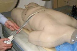
And it's easy to see why she feels that way. With volume sonography, ultrasound's benefits and capabilities can be significantly expanded, taking advantage of the ability to reconstruct volumes in many planes after a quick acquisition process, according to Benacerraf.
In her talk during a categorical course at the annual meeting of the American Institute of Ultrasound in Medicine (AIUM) last week in Orlando, Benacerraf shared Harvard research comparing 3D volume ultrasound with traditional 2D imaging for second-trimester fetal screening. The researchers found that 3D performed as well as 2D, and it was also much faster.
As an Ultrasound Insider subscriber, you have access to this Ultrasound Insider Exclusive story before it is published for the rest of our AuntMinnie.com members. To read more about the benefits of 3D, click here.
And be sure to check back in the Ultrasound Digital Community for continuing coverage of the AIUM meeting.
Have an idea for a topic you'd like to see covered, or an article you'd like to submit? As always, please feel free to drop me a line.







![Examples of ultrasound findings and techniques. (A) Images in a 39-year-old male patient with a mass in the left thigh. The mass is heterogeneous on the B-mode US image (compared with the patient in D) and showed increased microvascularity (superb microvascular imaging [SMI]) and shear-wave elastography (SWE) values. Undifferentiated pleomorphic sarcoma was diagnosed at biopsy (with pleomorphic rhabdomyosarcoma in surgical specimen). (B) Images in an 18-year-old male patient with a mass in the left leg. The mass is hypoechoic on the B-mode image, with no other findings suggestive of malignancy. The lesion is in contact with the cortex of the tibia, which is slightly irregular. CT revealed a doubtful anteromedial tibial erosion. The microvascular study demonstrated high vascularization, suggestive of malignancy. Periosteal Ewing sarcoma was diagnosed with both histologic and immunohistochemical confirmation. (C) Images in a 69-year-old female patient with a lump growing on the outside of the left leg. Multiple SWE examinations were performed (please note the high values obtained in the measurements, whereas the color map highlights the stiffness relative to adjacent tissues). SMI showed areas of increased vascularization to target for sampling. Undifferentiated spindle cell sarcoma was diagnosed at biopsy, with residual leiomyosarcoma in the surgical specimen after neoadjuvant therapy. (D) Images in a 56-year-old female patient with a mass in the right thigh. The mass is heterogeneous at both B-mode ultrasound (similar to patient A) and MRI (coronal T2-weighted spectral attenuated inversion recovery [SPAIR]; T1-weighted pre-contrast and postcontrast imaging), which even shows uptake after the administration of paramagnetic contrast material, which is traditionally suggestive of malignancy. Low values at SMI and elastography are suggestive of benignity. Spindle cell lipoma was diagnosed at biopsy, with atypical spindle cell lipomatous tumor in the surgical specimen.](https://img.auntminnie.com/mindful/smg/workspaces/default/uploads/2026/08/images-radiol250278fig2.APCFLSvX6p.jpg?auto=format%2Ccompress&fit=crop&h=112&q=70&w=112)


