Power Doppler sonography (PDS) has shown promise in detecting subclinical cases of juvenile rheumatoid arthritis (JRA) in a recent study. The technique was used along with grayscale ultrasound and clinical examination of symptoms to evaluate the therapeutic response in 16 girls and 14 boys with the condition in the knee joint.
Patients were treated with naproxen for six months, and assessed on three different occasions: on the day before treatment (baseline), at the end of two months of treatment, and at the end of six months of treatment. Clinical assessment, grayscale ultrasound, and PDS were done on each occasion.
PDS was used to assess the degree of vascularity in the knee joints, grayscale ultrasound was used to assess synovial effusion and thickness, and clinical assessment was carried out based on the presence of pain, arterial swelling, and functional impairment.
PDS detected three cases of mild hyperemia, while both clinical assessment and grayscale ultrasound showed no abnormality at the end of the sixth month of treatment. "PDS showed hyperemic changes in knees that were clinically normal," reported researchers from the Post Graduate Institute of Medical Education and Research (PGIMER) in Chandigarh. The group presented the study findings at the 2004 RSNA meeting in Chicago in December.
 |
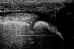
| PDS reveals normal vascularity. |
 |
| PDS shows markedly increased vascularity. |
In addition, PDS was able to demonstrate decreased vascularity in response to treatment in the second and sixth month. Vascularity, which was quantitatively assessed by grading the knee joints, decreased by one count in the second month and at least by one to two counts in the sixth month.
 |
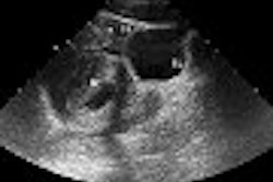
| Baseline PDS of right knee reveals marked vascular signals. |
 |
| Follow-up images of the same patient at the end of the second (above) and sixth (below) month after therapy demonstrate significant decrease in vascularity. |
 |
"PDS shows substantial promise in the evaluation of synovial inflammatory disease in subclinical cases of JRA, and is useful in objective assessment of therapeutic response and efficacy," said Dr Kushaljit Singh Sodhi, assistant professor at PGIMER, in his presentation at the meeting. His research colleagues were Drs C. Shanmugavel, S. Katariya, R. Sidhu, S. Singh, and Sudha Suri.
Ultrasound with power Doppler better
At the start of the study patients were classified into two groups: those with active knee involvement and those in clinical remission.
The classification was based on the total clinical score, which was computed by grading patients on three counts: pain (absent or present), articular swelling (absent, mild, moderate, or severe), and functional impairment (no restriction of movement, less than 5° extension, less than 10° extension, less than 15° extension, or greater than 15° extension). The individual scores were added to give the total clinical score.
All patients with a mean score greater than 1 were classified as having active knee involvement. Those with a score of 1 or lower were classified as being in clinical remission.
The two groups were scored depending on the degree of vascularity demonstrated on PDS performed in the suprapatellar region.
|
||||||||||||||||||||||||||||||||||||||||||
| Findings presented at RSNA 2004, courtesy of PGIMER. | ||||||||||||||||||||||||||||||||||||||||||
The researchers said the differential score between the two patient groups may reflect true variation in blood flow, depending on the disease activity. "If the disease activity is higher, blood flow will be more and the PDS score will be higher," Sodhi said.
The researchers also concluded that ultrasound with power Doppler is more sensitive in the detection and follow-up of clinically silent cases of JRA, based on a statistical analysis of the results obtained from grayscale ultrasound, PDS, and clinical assessment.
The Pearson co-efficient of correlation between the grayscale score and total clinical score and between the PDS score and total clinical score was calculated and compared. "Power Doppler shows more positive correlation than the grayscale score at follow-up in the second and sixth month," the researchers reported.
By N. Shivapriya
AuntMinnie.com staff writer
January 7, 2005
All images were presented at the 2004 RSNA meeting, and were provided courtesy of PGIMER.
Copyright © 2005 AuntMinnie.com