(Ultrasound Review) Radiologists at Einstein College of Medicine in New York City recently compared the sonographic appearances of the tubal ring of ectopic pregnancy with corpus luteum of the ovary.
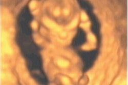
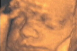
The tubal ring sign associated with an ectopic pregnancy consists of a thick-walled adnexal cystic structure shown separate to the ovary. When a pregnant patient presents with a similar structure adjacent to the ovary, but without clear separation, this could represent a corpus luteum or an ectopic pregnancy.
Given the mortality issues associated with ectopic pregnancy, Dr Marjorie Stein and colleagues sought to distinguish these two entities through a comparison of ultrasound findings. Results were published in Journal of Ultrasound in Medicine.
A retrospective review of first-trimester patients who had transvaginal ultrasound over a 20-month period revealed a cystic adnexal structure in 120 women with positive serum ß-human chorionic gonadotropin level. Seventy-nine women were included for study. Transvaginal imaging was performed using a 5 or 7 MHz, specialized transducer and color Doppler was employed.
"Each adnexal cystic structure was evaluated for six specific sonographic characteristics: the echogenicity of the wall of the cyst compared with that of the ovarian parenchyma and with that of the endometrial lining (excluding any fluid or debris), the average thickness of the cyst wall in two planes, the echo texture within the cystic cavity, flow distribution within the wall, and percentage of wall circumference with flow," they reported.
The adnexal mass was classified as clear when anechoic; lacy if multiple thin septations were present in a reticular pattern; solid if nonvascular soft-tissue was present; and ground-glass appearance if homogeneous low-level echoes were shown.
Color Doppler was used to determine blood flow distribution. Flow was categorized as absent; present within the wall; present within the full thickness of the wall from the inner to outer margins of the rind; and peripheral to the wall.
"Ancillary sonographic signs to distinguish between an ectopic pregnancy and a corpus luteum include decreased wall echogenicity compared with the endometrium and an anechoic texture, which suggests a corpus luteum," they concluded.
Although hemoperitoneum was not found in patients with corpora lutea, but was found in patients with ectopic pregnancy, it was not a specific finding, as hemoperitoneum also was found in patients with ruptured ovarian cyst.
Sonographic comparison of the tubal ring of ectopic pregnancy with the corpus luteumStein, M. W. et. al.
Albert Einstein College of Medicine and Montefiore Medical Center, New York City.
J Ultrasound Med 2004 January; 23:57-62
By Ultrasound Review
February 9, 2004
Copyright © 2004 AuntMinnie.com