Synaptive Medical has formed a collaborative clinical advisory board to support its long-term clinical and technology strategy across its surgical visualization, navigation, and MRI platforms.
According to the Toronto-based company, the Clinical Advisory Board, which comprises clinicians and surgeons, was created as a collaborative forum to contribute operating-room and clinical practice insight to inform platform evolution, clinical priorities, and evidence generation.
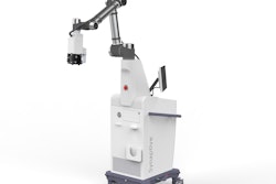
Synaptive develops technologies intended to aid in clinical decision-making for neurosurgery and related specialties, including an integrated suite spanning MRI, surgical planning, and robotic visualization.
The Clinical Advisory Board includes the following members:
- Constantinos Hadjipanayis, MD, PhD, of the University of Pittsburgh Medical Center and University of Pittsburgh School of Medicine
- G. Rees Cosgrove, MD, of Mass General Brigham and Harvard Medical School in Boston
- Elad Levy, MD, of the University at Buffalo in New York
- Nicholas Theodore, MD, of University Medical Center and Banner Health, both in Phoenix, AZ
- Dung Nguyen, MD, PharmD, of Stanford Health Care in Palo Alto, CA
- David Liebeskind, MD, of the University of Southern California (USC) Neurovascular Center and Keck School of Medicine and the University of California, Los Angeles (UCLA) School of Medicine
- Vitor Pereira, MD, of the University of Toronto
- Mitesh Shah, MD, of the Indiana University/Indiana University Health Neuroscience Institute and the Indiana University School of Medicine in Indianapolis


![Overview of the study design. (A) The fully automated deep learning framework was developed to estimate body composition (BC) (defined as subcutaneous adipose tissue [SAT] in liters; visceral adipose tissue [VAT] in liters; skeletal muscle [SM] in liters; SM fat fraction [SMFF] as a percentage; and intramuscular adipose tissue [IMAT] in deciliters) from MRI. The fully automated framework comprised one model (model 1) to quantify different BC measures (SAT, VAT, SM, SMFF, and IMAT) as three-dimensional (3D) measures from whole-body MRI scans. The second model (model 2) was trained to identify standardized anatomic landmarks along the craniocaudal body axis (z coordinate field), which allowed for subdividing the whole-body measures into different subregions typically examined on clinical routine MRI scans (chest, abdomen, and pelvis). (B) BC was quantified from whole-body MRI in over 66,000 individuals from two large population-based cohort studies, the UK Biobank (UKB) (36,317 individuals) and the German National Cohort (NAKO) (30,291 individuals). Bar graphs show age distribution by sex and cohort. BMI = body mass index. (C) After the performance assessment of the fully automated framework, the change in BC measures, distributions, and profiles across age decades were investigated. Age-, sex-, and height-adjusted body composition reference curves were calculated and made publicly available in a web-based z-score calculator (https://circ-ml.github.io).](https://img.auntminnie.com/mindful/smg/workspaces/default/uploads/2026/05/body-comp.XgAjTfPj1W.jpg?auto=format%2Ccompress&fit=crop&h=112&q=70&w=112)

