Cerebral "dirty-appearing" white matter (DAWM) -- an MRI finding considered a potential early marker of cerebrovascular disease -- is not associated with cognitive decline or dementia risk in healthy older adults, researchers have reported.
The findings could reframe how clinicians and researchers interpret this imaging abnormality, wrote a team led by doctoral candidate Ingmar Eiling of Leiden University Medical Center in the Netherlands. The group's results were presented at the recent International Society for Magnetic Resonance in Medicine (ISMRM) meeting and were published May 11 in Neurology.
"Baseline DAWM was not associated with cognition, cognitive decline, nor dementia risk in relatively healthy older adults," Eiling and colleagues noted. "DAWM may instead reflect early, subclinical pathology that has not yet reached a threshold sufficient to impair cognitive function."
DAWM refers to areas of diffuse signal hyperintensity visible on T2-weighted FLAIR MRI that fall between normal-appearing white matter and the more conspicuous white matter hyperintensities associated with cerebral small vessel disease (cSVD). Because cSVD has been associated with accelerated cognitive decline and increased dementia risk, the presence of DAWM has attracted interest among researchers as a possible early imaging biomarker. Clarifying whether DAWM carries independent prognostic value for cognition could improve how radiologists characterize and report these findings and help them better stratify patient risk.
"[The study was difficult] because DAWM is not easy to reliably detect," Eiling told AuntMinnie. "That's part of the reason why we started rating it visually in thousands of scans of older adults, to maximize the chance of finding some link to dementia later on."
Eiling and colleagues conducted a study that included data from the Age, Gene/Environment Susceptibility (AGES) Reykjavik longitudinal cohort, a population-based study comprised of more than 1% of Iceland's population. They selected 2,081 participants born between 1907 and 1935 with limited baseline white matter hyperintensities burden.
Two blinded observers visually rated DAWM on 1.5 Tesla FLAIR MRI scans as a percentage of lobar white matter volume (0%; 0% to 10%; 10% to 25%; and more than 25%). Study participants also underwent cognitive assessments at study baseline and at five-year follow-up that evaluated their memory, executive function, and processing speed; the researchers assessed patients' dementia status at 10-year follow-up.
The overall finding the team reported was that DAWM ratings were not associated with baseline cognitive scores across any domain or with cognitive decline at five years. They were also not associated with increased dementia risk at 10 years. However, baseline white matter hyperintensity volume was linked to lower executive function and processing speed scores, five-year decline in processing speed, and a 35% higher dementia risk over 10 years.
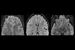
 Examples of DAWM on FLAIR MRI slices. Transversal slices of 1.5T FLAIR MRI scans. For visualization only, brains were skull-stripped and DAWM is shown with dotted lines (occipital DAWM is red, parietal DAWM is orange). Top row images (25% of lobar WM) show larger, more confluent configurations of DAWM, extending beyond the slices shown here.Ingmar Eiling and ISMRM
The study results don't diminish the relevance of DAWM, but instead point to where research must go next, according to Eiling.
"Clinically, [the presence of DAWM is an] understudied part of [MRI] scans that have been available for over 25 years," he said. "Perhaps the key clinical relevance of this study is that subtle brain changes will not always have clear implications for later dementia risk."
Access the Neurology paper here and our full coverage of ISMRM here.