Signs of injury to the brain's white matter called white matter hyperintensities, as seen on brain scans, may be tied more strongly to vascular risk factors, brain shrinkage, and other markers of dementia in former tackle football players than in those who did not play football.
The results add to previous research that has evaluated the effect of repeated head injuries, senior author Michale Alosco, PhD, of Boston University said in a statement released by the journal. The findings were published December 20 in the journal Neurology.
"Studies have shown that athletes exposed to repetitive head impacts can have increased white matter hyperintensity burden in their brains," he said. "White matter hyperintensities are easily seen on MRI as markers of injury of various causes. We know these markers are more common as people age and with medical conditions such as high blood pressure, but [our] results provide initial insight that they may be related to multiple aspects of brain damage from repetitive head impacts."
Tracking levels of white matter hyperintensities on brain MRI exams shows promise for evaluating long-term effects of repetitive head impacts, Alosco and colleagues noted. Repetitive head impacts have also been linked to chronic traumatic encephalopathy (CTE), a neurodegenerative disease that can result in dementia, they explained.
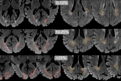
The investigators conducted a study that included 120 former professional football players and 60 former college football players (average age, 57). They then compared this group to a cohort of 60 men (average age, 59) who had no symptoms, did not play football, and had no history of repetitive head impacts or concussion. Each of the study participants underwent brain MRI scans and lumbar punctures to identify any biomarkers of neurodegenerative disease and white matter changes.
Alosco's group found the following:
- In the former football players, a higher level of white matter hyperintensities was linked to greater vascular risk factors; higher concentrations of p-tau proteins found in Alzheimer's disease, CTE, and other neurodegenerative diseases; more brain shrinkage; and a decrease in the health of the white matter pathways in the brain.
- The relationship between white matter hyperintensities and stroke risk was 11 times stronger in former football players than in those who did not play football, 2.5 times stronger for the concentration of p-tau proteins, and four times stronger for white matter integrity.
The study does not prove that repetitive head impacts and white matter hyperintensities cause other brain changes, but only shows an association, the group noted.
The complete study can be found here.


![Overview of the study design. (A) The fully automated deep learning framework was developed to estimate body composition (BC) (defined as subcutaneous adipose tissue [SAT] in liters; visceral adipose tissue [VAT] in liters; skeletal muscle [SM] in liters; SM fat fraction [SMFF] as a percentage; and intramuscular adipose tissue [IMAT] in deciliters) from MRI. The fully automated framework comprised one model (model 1) to quantify different BC measures (SAT, VAT, SM, SMFF, and IMAT) as three-dimensional (3D) measures from whole-body MRI scans. The second model (model 2) was trained to identify standardized anatomic landmarks along the craniocaudal body axis (z coordinate field), which allowed for subdividing the whole-body measures into different subregions typically examined on clinical routine MRI scans (chest, abdomen, and pelvis). (B) BC was quantified from whole-body MRI in over 66,000 individuals from two large population-based cohort studies, the UK Biobank (UKB) (36,317 individuals) and the German National Cohort (NAKO) (30,291 individuals). Bar graphs show age distribution by sex and cohort. BMI = body mass index. (C) After the performance assessment of the fully automated framework, the change in BC measures, distributions, and profiles across age decades were investigated. Age-, sex-, and height-adjusted body composition reference curves were calculated and made publicly available in a web-based z-score calculator (https://circ-ml.github.io).](https://img.auntminnie.com/mindful/smg/workspaces/default/uploads/2026/05/body-comp.XgAjTfPj1W.jpg?auto=format%2Ccompress&fit=crop&h=100&q=70&w=100)


![Overview of the study design. (A) The fully automated deep learning framework was developed to estimate body composition (BC) (defined as subcutaneous adipose tissue [SAT] in liters; visceral adipose tissue [VAT] in liters; skeletal muscle [SM] in liters; SM fat fraction [SMFF] as a percentage; and intramuscular adipose tissue [IMAT] in deciliters) from MRI. The fully automated framework comprised one model (model 1) to quantify different BC measures (SAT, VAT, SM, SMFF, and IMAT) as three-dimensional (3D) measures from whole-body MRI scans. The second model (model 2) was trained to identify standardized anatomic landmarks along the craniocaudal body axis (z coordinate field), which allowed for subdividing the whole-body measures into different subregions typically examined on clinical routine MRI scans (chest, abdomen, and pelvis). (B) BC was quantified from whole-body MRI in over 66,000 individuals from two large population-based cohort studies, the UK Biobank (UKB) (36,317 individuals) and the German National Cohort (NAKO) (30,291 individuals). Bar graphs show age distribution by sex and cohort. BMI = body mass index. (C) After the performance assessment of the fully automated framework, the change in BC measures, distributions, and profiles across age decades were investigated. Age-, sex-, and height-adjusted body composition reference curves were calculated and made publicly available in a web-based z-score calculator (https://circ-ml.github.io).](https://img.auntminnie.com/mindful/smg/workspaces/default/uploads/2026/05/body-comp.XgAjTfPj1W.jpg?auto=format%2Ccompress&fit=crop&h=112&q=70&w=112)


