An international panel of 21 specialists has issued consensus recommendations for using MRI in prostate cancer screening, offering detailed guidance on how scans should be acquired, interpreted, and reported.
A team led by Nikhil Mayor, MBBS, of Imperial College London in the U.K., wrote the Prostate Imaging Standards for Screening MRI (PRISM) guidance to "provide consensus on the acquisition, interpretation, and reporting of prostate MRI for cancer screening." The document was published June 11 in JAMA Oncology.
PRISM emerged from a review of six studies found on PubMed, Cochrane Central Register of Controlled Trials (CENTRAL), Scopus, Web of Science, and ClinicalTrials.gov in September 2024. The studies involved 1,426 men who underwent screening MRI; PRISM authors combined these results with a structured consensus exercise conducted by a panel of eight urologists, 11 radiologists, and two pathologists from six countries. Of 323 consensus statements, 235 (72.8%) reached agreement among panel members.
The guidance calls for prostate cancer screening to begin at age 50 and continue through age 70 for most men, with Black men invited from age 45 given their elevated risk. Men with a life expectancy under 10 years should not be screened, and those with a family history of the disease or high-risk genetic variants should be offered earlier entry into screening programs.
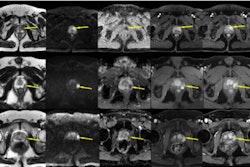
The authors recommended that MR imaging for this indication should follow a prostate-specific antigen (PSA) blood test rather than be performed as a standalone first step, although they did not reach consensus on the exact PSA threshold that should trigger an MRI scan. They recommended that the MRI protocol be kept to 15 minutes and should use only T2-weighted and diffusion-weighted imaging without contrast injection or endorectal coils.
They also suggested a "stage-gated" reading approach, under which radiologists review only axial T2-weighted and high b-value diffusion images, moving on to interpret the full scan only if a suspicious lesion appears on both sequences simultaneously. This approach has been shown to halve biopsy recommendation rates while also improving the proportion of scans that correctly identify clinically significant cancer, according to the team.
Access the full guideline here.