TORONTO - A relatively new ultrafast MRI protocol called wave-CAIPI cuts diagnostic scan time for dementia by 66%, according to research presented June 5 at the International Society for Magnetic Resonance in Medicine (ISMRM) meeting.
The findings could definitely improve patient care, presenter Haroon Chughtai, PhD, of University College London in the U.K. and colleagues noted.
"Our ultrafast protocol enabled by Wave-CAIPI shows promise in reducing the diagnostic scan time for dementia from around 18 minutes to under six minutes while retaining clinical utility across several contrasts," they wrote.
Structural brain imaging is an important tool for diagnosing cognitive disorders and dementias, including identifying Alzheimer's disease, Chughtai noted. MRI is the go-to imaging modality for this indication because it can image soft tissue, but its use can be hindered by long scan times, high cost, and less availability compared to CT. That's why shorter scan times could enable wider access to MRI as a first-line modality for cognitive disease.
"[For diagnosis of cognitive disease] patients are often imaged with CT rather than MRI, generating less detailed scans and leading to variations in diagnosis accuracy and disease management," Chughtai told session attendees. "[And] with the imminent arrival of disease-modifying therapies for Alzheimer's disease, demand for diagnostic MRI is set to increase substantially ... [so that] reducing scan times improves availability by making MRIs easier to schedule, more affordable, and easier to tolerate."
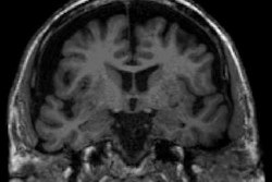
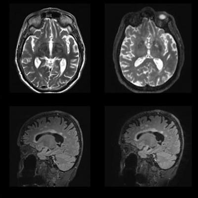
 Examples of scans acquired from the clinical and rapid protocols for each sequence. Image and caption courtesy of Haroon Chughtai, PhD.
MRI for diagnosing dementia does have some limits, most notably image degradation that can affect the clinical usefulness of its images, according to Chughtai. To address this limitation, his group developed a prototype ultrafast MRI protocol based on a technique called wave-controlled aliasing in parallel imaging, or CAIPI, which is designed to speed 3D exams.
To evaluate this protocol, the team conducted a study that included 40 patients recruited from a cognitive disorders clinic at the National Hospital for Neurology and Neurosurgery in London. All underwent a 3-tesla MRI exam (Magnetom Prisma, Siemens Healthineers) as part of their diagnostic workup.
The standard protocol consisted of T1w, T2w, FLAIR and T2*/SWI sequences, while the rapid protocol used Siemens' work-in-progress wave-CAIPI sequences (3D MPRAGE, 3D FLAIR, 3D T2 and 3D T2*).
A team of six readers consisting of two neuroradiologists, two physicists, and two neurologists read the exams, scoring them on a three-point scale of -1 to +1 (with -1 equal to a preference for the standard of care MRI sequence and +1 equal to a preference for the rapid sequence).
The study found that the rapid protocol reduced scan time by 66%, from 17 minutes 39 seconds to 5 minutes 20 seconds.
The investigators also reported the following:
- Across all comparisons the raters performed for T1w sequences, they deemed 90% of the rapid scans as equivalent or superior to the standard protocol.
- They were less enthusiastic about the rapid 3D T2 and 3D T2* sequence, judging the rapid 3D T2 inferior "due to coarser in-plane axial resolution hindering identification of perivascular spaces."
- Across all comparisons the raters performed for the rapid FLAIR sequence, 65% of readers assessed them to be equivalent or superior -- although two readers decreased their rating due to "reduced image sharpness."
The findings show promise for better dementia diagnosis, Chughtai and colleagues concluded.
"Our preliminary results ... demonstrate a substantial reduction in scan times, leading to potential cost-savings for providers and better care for patients," they wrote. "Further improvements in data acquisition and image processing may yet yield a better balance of speed, clinical utility, and image quality."