Image analysis software developer Perspectum Diagnostics highlighted two studies presented during the American Association for the Study of Liver Diseases (AASLD) Liver Meeting held November 9-13.
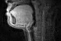
The abstracts detailed the use of Perspectum's technology for improving upon MR cholangiopancreatography (MRCP). MRCP is a noninvasive technique used in the evaluation of biliary tree anatomy, but it is not quantitative and has high subjectivity in reading.
Perspectum's MRCP+, on the other hand, provides information that increases diagnostic confidence while minimizing the frequency of invasive procedures, according to the company. MRCP+ is designed to allow existing MRCP data to be enhanced and quantitatively characterized using advanced image processing techniques. The software enhances data from conventional MRCP without the need for a contrast agent.
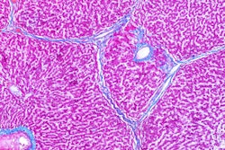
In one study, researchers evaluated the potential clinical utility of the metrics produced by MRCP+ for discriminating biliary disease. Heavily T2-weighted MRCP images were acquired in a cohort of healthy subjects, as well as those with autoimmune hepatitis, hepatitis C, nonalcoholic fatty liver disease, primary biliary cholangitis, and primary sclerosing cholangitis. The images were processed with MRCP+ to enhance and quantify the tubular biliary structures.
MRCP+ produced reference intervals of healthy common bile duct width that were comparable to those previously reported in the literature, and patients with primary sclerosing cholangitis had a significantly higher duct variability score than other cohorts. Demonstrating that a multiscale image analysis method both enhanced and quantified biliary tubular structures, the results show that MRCP+ provides measures that could objectively differentiate patients with primary sclerosing cholangitis.
In another study, matrix metalloproteinase 7 (MMP7) was researched as a novel biomarker in pediatric sclerosing cholangitis. MRCP+ was used to determine the percentage of biliary tree affected by strictures and dilatations.
This study showed, with good correlation to MRCP+, that MMP7 is a promising biomarker for the noninvasive diagnosis of primary sclerosing cholangitis in children.
MRCP+ is pending clearance from the U.S. Food and Drug Administration (FDA).


![Overview of the study design. (A) The fully automated deep learning framework was developed to estimate body composition (BC) (defined as subcutaneous adipose tissue [SAT] in liters; visceral adipose tissue [VAT] in liters; skeletal muscle [SM] in liters; SM fat fraction [SMFF] as a percentage; and intramuscular adipose tissue [IMAT] in deciliters) from MRI. The fully automated framework comprised one model (model 1) to quantify different BC measures (SAT, VAT, SM, SMFF, and IMAT) as three-dimensional (3D) measures from whole-body MRI scans. The second model (model 2) was trained to identify standardized anatomic landmarks along the craniocaudal body axis (z coordinate field), which allowed for subdividing the whole-body measures into different subregions typically examined on clinical routine MRI scans (chest, abdomen, and pelvis). (B) BC was quantified from whole-body MRI in over 66,000 individuals from two large population-based cohort studies, the UK Biobank (UKB) (36,317 individuals) and the German National Cohort (NAKO) (30,291 individuals). Bar graphs show age distribution by sex and cohort. BMI = body mass index. (C) After the performance assessment of the fully automated framework, the change in BC measures, distributions, and profiles across age decades were investigated. Age-, sex-, and height-adjusted body composition reference curves were calculated and made publicly available in a web-based z-score calculator (https://circ-ml.github.io).](https://img.auntminnie.com/mindful/smg/workspaces/default/uploads/2026/05/body-comp.XgAjTfPj1W.jpg?auto=format%2Ccompress&fit=crop&h=100&q=70&w=100)


![Overview of the study design. (A) The fully automated deep learning framework was developed to estimate body composition (BC) (defined as subcutaneous adipose tissue [SAT] in liters; visceral adipose tissue [VAT] in liters; skeletal muscle [SM] in liters; SM fat fraction [SMFF] as a percentage; and intramuscular adipose tissue [IMAT] in deciliters) from MRI. The fully automated framework comprised one model (model 1) to quantify different BC measures (SAT, VAT, SM, SMFF, and IMAT) as three-dimensional (3D) measures from whole-body MRI scans. The second model (model 2) was trained to identify standardized anatomic landmarks along the craniocaudal body axis (z coordinate field), which allowed for subdividing the whole-body measures into different subregions typically examined on clinical routine MRI scans (chest, abdomen, and pelvis). (B) BC was quantified from whole-body MRI in over 66,000 individuals from two large population-based cohort studies, the UK Biobank (UKB) (36,317 individuals) and the German National Cohort (NAKO) (30,291 individuals). Bar graphs show age distribution by sex and cohort. BMI = body mass index. (C) After the performance assessment of the fully automated framework, the change in BC measures, distributions, and profiles across age decades were investigated. Age-, sex-, and height-adjusted body composition reference curves were calculated and made publicly available in a web-based z-score calculator (https://circ-ml.github.io).](https://img.auntminnie.com/mindful/smg/workspaces/default/uploads/2026/05/body-comp.XgAjTfPj1W.jpg?auto=format%2Ccompress&fit=crop&h=112&q=70&w=112)


