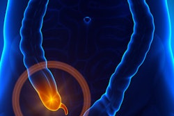
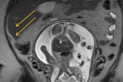
Ultrasound, CT, and MRI are all viable supplemental imaging modalities for assessing acute appendicitis after initial ultrasound, according to a study published online June 19 in Radiology.
The team led by Dr. Kevin Eng conducted a literature search on Medline and Embase, identifying studies that used surgery or histopathologic exam information alone or in combination with clinical follow-up or chart review to assess the diagnostic accuracy of supplemental imaging modalities for appendicitis.
Studies included the following:
- For children -- ultrasound: six studies and 548 patients; CT: nine studies and 1,498 patients; MRI: five studies and 287 patients
- For adults -- ultrasound: three studies and 169 patients; CT: 11 studies and 1,027 patients; MRI: six studies and 427 patients
All three modalities had comparable, high accuracy for diagnosing the disease, both in children and adults, the researchers found.
| Pooled sensitivities and specificities of second-line imaging for diagnosing appendicitis | |||
| Measure | Ultrasound | CT | MRI |
| Sensitivity: Adults | 83.1% | 89.9% | 97.4% |
| Sensitivity: Children | 91.3% | 96.2% | 89.9% |
| Specificity: Adults | 90.9% | 93.6% | 97.1% |
| Specificity: Children | 95.2% | 94.6% | 93.6% |
"All three modalities may be valid as second-line imaging in a clinical imaging pathway for diagnosis and management of appendicitis," they concluded.