
When researchers recreated an anterior cruciate ligament (ACL) rupture using 3D models based on MR scans of knees, they found no mechanistic differences in the injury between men and women, according to an article published online April 18 in the American Journal of Sports Medicine.
Previous studies have suggested that the higher incidence of ACL tears in women than men -- the injury is two to four times more common in women -- is a consequence of inherent differences in knee movement, noted senior author Louis DeFrate, PhD, and colleagues at Duke University Medical Center. The findings primarily came from reviewing slow-motion replays of ACL injuries as they occurred.
"Watching videos of athlete injuries, previous researchers have suggested that females may have a different mechanism of injury than males," DeFrate said in a statement from the university. "But it's difficult to determine the precise position of the knee and the time of injury through footage."
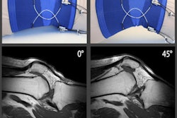
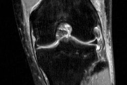
Favoring a more in-depth approach, the investigators examined ACL tears evident on the knee MR images of 15 men and 15 women, acquired within one month after the injury. They identified a common bruising pattern on the surfaces of the femur and tibia -- two large bones that collide after an ACL ruptures.
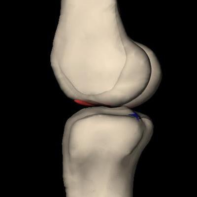
Applying this information to computer algorithms, the group was able to create 3D models of knee MR images for visualizing the position of the knee bones during injury. The researchers concluded that knee positioning for an ACL tear was the same in men and women.
3D reconstruction of an ACL tear based on MRI scans, computer algorithms, and 3D modeling. Image courtesy of Louis DeFrate, PhD, at Duke University.This finding is the latest from a series of studies focused on understanding ACL injuries that DeFrate and colleagues began in 2008. They have since published research proposing that ACL tears are caused by landing on a hyperextended knee, not the inward buckling motion of the knee that often follows the injury. DeFrate received the 2016 Kappa Delta Young Investigator Award from the American Academy of Orthopaedic Surgeons for his work in the field.
"In order to develop improved treatment strategies and prevention, we need a clear understanding of what motions are most dangerous for athletes," he said. "This work provides new evidence that landing on an extended knee may be a dangerous position for ACL tears in both males and females."


![Overview of the study design. (A) The fully automated deep learning framework was developed to estimate body composition (BC) (defined as subcutaneous adipose tissue [SAT] in liters; visceral adipose tissue [VAT] in liters; skeletal muscle [SM] in liters; SM fat fraction [SMFF] as a percentage; and intramuscular adipose tissue [IMAT] in deciliters) from MRI. The fully automated framework comprised one model (model 1) to quantify different BC measures (SAT, VAT, SM, SMFF, and IMAT) as three-dimensional (3D) measures from whole-body MRI scans. The second model (model 2) was trained to identify standardized anatomic landmarks along the craniocaudal body axis (z coordinate field), which allowed for subdividing the whole-body measures into different subregions typically examined on clinical routine MRI scans (chest, abdomen, and pelvis). (B) BC was quantified from whole-body MRI in over 66,000 individuals from two large population-based cohort studies, the UK Biobank (UKB) (36,317 individuals) and the German National Cohort (NAKO) (30,291 individuals). Bar graphs show age distribution by sex and cohort. BMI = body mass index. (C) After the performance assessment of the fully automated framework, the change in BC measures, distributions, and profiles across age decades were investigated. Age-, sex-, and height-adjusted body composition reference curves were calculated and made publicly available in a web-based z-score calculator (https://circ-ml.github.io).](https://img.auntminnie.com/mindful/smg/workspaces/default/uploads/2026/05/body-comp.XgAjTfPj1W.jpg?auto=format%2Ccompress&fit=crop&h=100&q=70&w=100)


![Overview of the study design. (A) The fully automated deep learning framework was developed to estimate body composition (BC) (defined as subcutaneous adipose tissue [SAT] in liters; visceral adipose tissue [VAT] in liters; skeletal muscle [SM] in liters; SM fat fraction [SMFF] as a percentage; and intramuscular adipose tissue [IMAT] in deciliters) from MRI. The fully automated framework comprised one model (model 1) to quantify different BC measures (SAT, VAT, SM, SMFF, and IMAT) as three-dimensional (3D) measures from whole-body MRI scans. The second model (model 2) was trained to identify standardized anatomic landmarks along the craniocaudal body axis (z coordinate field), which allowed for subdividing the whole-body measures into different subregions typically examined on clinical routine MRI scans (chest, abdomen, and pelvis). (B) BC was quantified from whole-body MRI in over 66,000 individuals from two large population-based cohort studies, the UK Biobank (UKB) (36,317 individuals) and the German National Cohort (NAKO) (30,291 individuals). Bar graphs show age distribution by sex and cohort. BMI = body mass index. (C) After the performance assessment of the fully automated framework, the change in BC measures, distributions, and profiles across age decades were investigated. Age-, sex-, and height-adjusted body composition reference curves were calculated and made publicly available in a web-based z-score calculator (https://circ-ml.github.io).](https://img.auntminnie.com/mindful/smg/workspaces/default/uploads/2026/05/body-comp.XgAjTfPj1W.jpg?auto=format%2Ccompress&fit=crop&h=112&q=70&w=112)


