Tuesday, November 27 | 11:00 a.m.-11:10 a.m. | SSG11-04 | Room E450B
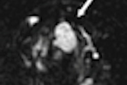
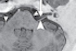
Chinese researchers have found that a 3D fast imaging with steady-state acquisition (FIESTA) MR imaging sequence can significantly enhance the identification of the anterior cruciate ligament (ACL) compared with sagittal spin-echo T1-weighted MRI.In addition, the rotating curved planar reconstruction of the 3D FIESTA sequence can improve the delineation of the entire ACL, according to lead study author Dr. Dapeng Hao, PhD, from the Affiliated Hospital of Qingdao University Medical College, and colleagues.
Hao and colleagues took 3-tesla MRI exams of 40 knee joints in healthy volunteers using a phased-array extremity coil. The imaging protocol included oblique sagittal spin-echo T1-weighted MRI and the 3D FIESTA sequence. The rotating curved-planar reconstructions of the ACL were conducted at 0°, 30°, 60°, 90°, 120°, 150°, and 180°.
Signal-to-noise and contrast-to-noise ratios with 3D FIESTA were compared with those of oblique sagittal spin-echo T1-weighted MRI. The researchers also rated on a three-point scale the presence of the tibial attachment, femoral attachment, and double bundles of the ACL on oblique sagittal spin-echo T1-weighted and curved planar reconstructions of 3D FIESTA MRI.
Their analysis found that the ACL signal-to-noise ratio efficiency of the 3D FIESTA sequence was significantly greater than that of the oblique spin-echo T1-weighted MRI sequence.
3D FIESTA also produced images with significantly greater contrast-to-noise ratios between ACL and synovial fluid than the oblique spin-echo T1-weighted MRI. Curved planar reconstruction by 3D FIESTA MRI also provided excellent visualization of the ACL.


![Overview of the study design. (A) The fully automated deep learning framework was developed to estimate body composition (BC) (defined as subcutaneous adipose tissue [SAT] in liters; visceral adipose tissue [VAT] in liters; skeletal muscle [SM] in liters; SM fat fraction [SMFF] as a percentage; and intramuscular adipose tissue [IMAT] in deciliters) from MRI. The fully automated framework comprised one model (model 1) to quantify different BC measures (SAT, VAT, SM, SMFF, and IMAT) as three-dimensional (3D) measures from whole-body MRI scans. The second model (model 2) was trained to identify standardized anatomic landmarks along the craniocaudal body axis (z coordinate field), which allowed for subdividing the whole-body measures into different subregions typically examined on clinical routine MRI scans (chest, abdomen, and pelvis). (B) BC was quantified from whole-body MRI in over 66,000 individuals from two large population-based cohort studies, the UK Biobank (UKB) (36,317 individuals) and the German National Cohort (NAKO) (30,291 individuals). Bar graphs show age distribution by sex and cohort. BMI = body mass index. (C) After the performance assessment of the fully automated framework, the change in BC measures, distributions, and profiles across age decades were investigated. Age-, sex-, and height-adjusted body composition reference curves were calculated and made publicly available in a web-based z-score calculator (https://circ-ml.github.io).](https://img.auntminnie.com/mindful/smg/workspaces/default/uploads/2026/05/body-comp.XgAjTfPj1W.jpg?auto=format%2Ccompress&fit=crop&h=100&q=70&w=100)


![Overview of the study design. (A) The fully automated deep learning framework was developed to estimate body composition (BC) (defined as subcutaneous adipose tissue [SAT] in liters; visceral adipose tissue [VAT] in liters; skeletal muscle [SM] in liters; SM fat fraction [SMFF] as a percentage; and intramuscular adipose tissue [IMAT] in deciliters) from MRI. The fully automated framework comprised one model (model 1) to quantify different BC measures (SAT, VAT, SM, SMFF, and IMAT) as three-dimensional (3D) measures from whole-body MRI scans. The second model (model 2) was trained to identify standardized anatomic landmarks along the craniocaudal body axis (z coordinate field), which allowed for subdividing the whole-body measures into different subregions typically examined on clinical routine MRI scans (chest, abdomen, and pelvis). (B) BC was quantified from whole-body MRI in over 66,000 individuals from two large population-based cohort studies, the UK Biobank (UKB) (36,317 individuals) and the German National Cohort (NAKO) (30,291 individuals). Bar graphs show age distribution by sex and cohort. BMI = body mass index. (C) After the performance assessment of the fully automated framework, the change in BC measures, distributions, and profiles across age decades were investigated. Age-, sex-, and height-adjusted body composition reference curves were calculated and made publicly available in a web-based z-score calculator (https://circ-ml.github.io).](https://img.auntminnie.com/mindful/smg/workspaces/default/uploads/2026/05/body-comp.XgAjTfPj1W.jpg?auto=format%2Ccompress&fit=crop&h=112&q=70&w=112)


