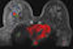
(Booth 423) Breast MRI technology developer Aurora Imaging Technology of North Andover, MA, will unveil AuroraEdge, a new image-enhancement technique for its 1.5-tesla Aurora scanner.

The feature is designed to counteract aliasing or wrap-around artifacts that are produced when areas outside the field-of-view are folded back into the image. The problem has become more severe with the adaptation of parallel imaging on whole-body MRI systems, according to the company. AuroraEdge oversamples in both in-plane dimensions to avoid aliasing in those dimensions.
In addition, motion artifacts from cardiac and respiratory movement often impair visualization of the axillary tail of the breast and axillary regions on most whole-body MRI scans, the company said. Motion also can affect the quality of breast imaging. The pulse sequence and image-acquisition system used in AuroraEdge minimizes the effect of cardiac and respiratory motion on the images.
Image-acquisition specifications for AuroraEdge include a 3D 360 x 360 x 180 image-acquisition matrix and a 3D 512 x 512 x 250-slice reconstruction matrix. Slice thickness is 0.7 mm, with no gap between slices, and acquisition is isotropic in all three planes. Images are acquired in less than three minutes per scan, with 18 minutes total patient scan time.