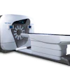
Belgium-based medtech company Xeos highlighted a case report published in Head & Neck describing the use of its Aura 10 mobile intraoperative PET/CT system in a patient with recurrent oral tongue squamous cell carcinoma.
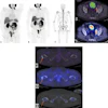
Investigators from Vanderbilt University Medical Center in Nashville, TN, used the system to perform specimen imaging immediately following resection in a 79-year-old woman who had received neoadjuvant chemoimmunotherapy prior to surgery. Specimen PET/CT revealed two separate residual tumor foci divided by an 8-mm bridge of inflammation without viable cancer, findings that closely correlated with subsequent pathology results, according to the report.
Intraoperative specimen imaging may help bridge the gap between presurgical imaging and residual disease identified on final pathology, Xeos said.
The case report can be found here.