The Society of Nuclear Medicine and Molecular Imaging (SNMMI) has highlighted a study showing that biopsies guided by prostate-specific membrane antigen (PSMA) PET/CT detect more prostate cancer than standard systematic biopsies in men with negative MRI scans.
The findings suggest that PSMA-PET/CT could provide an additional tool for identifying men who remain at risk despite nondiagnostic MRI findings and for directing the biopsy toward the most suspicious area, according to study lead Andrea Farolfi, MD, of the IRCCS Azienda Ospedaliero-Universitaria di Bologna in Italy.
“A negative or unclear MRI does not completely rule out clinically significant prostate cancer. As a result, some men may undergo repeated or extensive biopsies, while relevant tumors may still be missed,” Farolfi said, in an October 5 SNMMI news release.
Multiparametric MRI is considered the cornerstone for detecting and localizing clinically significant prostate cancer and enabling targeted biopsy, yet up to 20 to 30 percent of clinically significant prostate cancer cases may be missed in patients with negative or inconclusive MRI findings, SNMMI noted.
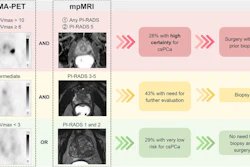
To determining whether PSMA-PET can refine risk stratification and guide targeted sampling for these men, Farolfi and colleagues recruited 63 men with negative or inconclusive MRI scans. Participants underwent gallium-68 PSMA-11 PET/CT followed by PET fusion-guided prostate biopsy. The researchers assessed the diagnostic performance of PSMA-PET/CT–guided targeted biopsy versus systematic biopsy.
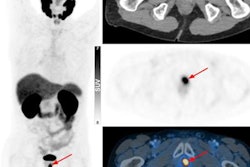
 Upstage with PET-guided biopsy. A 70-y-old patient with suspicious prostate cancer (PI-RADS 2; initial PSA, 15.9; PSA density, 0.4). Exam showed intense focal gallium-68 PSMA-11 uptake (SUVmax, 9.5; PRIMARY score 4) in left peripheral zone at midlevel shown maximum-intensity projection (A), transaxial view of CT image (B), PET image (C), and transaxial fused PET/CT image (D). Transperineal PSMA-PET targeted prostate biopsy revealed clinically significant prostate cancer (csPCa), ISUP 5, whereas standard biopsy revealed csPCa, ISUP 2. SNMMI
Ultimately, the results support the inclusion of PSMA-PET/CT in diagnostic pathways for this challenging cohort, and if confirmed in larger studies, the approach could make the diagnostic pathway more accurate and personalized, Farolfi said.
The full study was published in the September issue of the Journal of Nuclear Medicine and is available here.