Diffusion MRI yields promise for estimating the risk of progression from mild cognitive impairment to dementia, suggest findings to be presented May 13 at the International Society for Magnetic Resonance in Medicine (ISMRM) annual meeting in Honolulu, HI.
A team led by Aziz Ulug, PhD, from the University of Canterbury in New Zealand found that progression to dementia was significantly tied to less restricted diffusion and increased free fluid signal at baseline, noting that this may reflect cell loss. The group also found that increased diffusivity was linked to higher risk of progression.
“The regions with the highest sensitivity have been previously associated with Alzheimer’s disease, and show changes consistent with neuronal loss,” the Ulug team wrote.
 The hazard ratio for mean diffusivity and reciprocal of the hazard ratio for fractional anisotropy. Increases in mean diffusivity and decreases in fractional anisotropy are associated with a higher risk of conversion.ISMRM
Alzheimer’s researchers have made strides in understanding the mechanisms behind the disease. With the recent approval of Alzheimer's drugs, there is a need for better stratification of patients who may benefit from early interventions, the authors noted.
Using diffusion imaging with a multicompartment model, Ulug and colleagues sought to predict which patients with mild cognitive impairment will progress to Alzheimer’s via a study that included 40 participants classified with mild cognitive impairment at their first imaging session. Of these, 10 progressed to dementia.
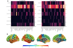
 Correlations amongst regions between hazard ratios derived from different dMRI metrics. The decreased restricted isotropic and increased free fluid components in the posterior cingulate are strongly associated with the risk of conversion to AD (1/HR=9.9, HR=4.34 respectively), as is the hindered compartment in the temporal pole (1/HR=4.87). n0s1=Restricted isotropic, n0s2=Hindered isotropic, n0s3=Free fluid, nds1=Restristced directional, MD=Mean diffusivity, FA=Fractional anisotropy.ISMRM
The investigators found that the following regions had the most significant effect sizes:
- Restricted isotropic compartment in the hippocampus (ꞵ = -0.946, p = 0.0078) and the posterior cingulate (ꞵ = -2.296, p = 0.0012)
- The free fluid component in the caudal anterior cingulate (ꞵ = 1.304, p = 0.0044) and posterior cingulate (ꞵ = 1.468, p = 0.0037)
- The hindered isotropic component in the temporal pole (ꞵ = -1.583, p = 0.0092)
Of the conventional diffusion tensor imaging metrics, the team reported statistical significance for the average diffusivity in the caudal anterior cingulate (ꞵ = 1.240, p = 0.0072) and posterior cingulate (ꞵ = 1.634, p = 0.0017).
The authors called for larger studies with longer follow-up appointments to help further show whether these results can provide valuable clinical information to stratify patients who may be optimal candidates for early treatment interventions. They also suggested that diffusion imaging-derived maps that are sensitive to tissue microstructures could serve as a biomarker to predict conversion.
Check out AuntMinnie.com’s full coverage of ISMRM 2025 here.