An FDG-PET/CT scan and thyroid biopsy changed a patient's diagnosis from terminal to treatable with the discovery of a rare neurosarcoidosis, according to a case published August 28 in Radiology Case Reports.
"Before undergoing this evaluation, the patient was given a terminal diagnosis and recommended hospice," wrote lead author Catherine Bullock, MD, of the Mayo Clinic Florida in Jacksonville, and colleagues.
Sarcoidosis is an inflammatory disease characterized by the formation of granulomas (tiny clusters of white blood cells), most commonly in the lungs, lymph nodes, skin, and liver. Less commonly, it can affect the central or peripheral nervous system, with a wide range of clinical manifestations.
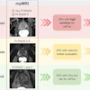
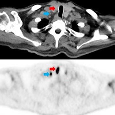
 Selected images from the initial FDG-PET/CT. Coronal FDG PET maximum intensity projection (MIP; A) demonstrates multifocal FDG avid foci including a right thyroid nodule (red arrow), multiple supraclavicular, mediastinal, hilar, and mesenteric lymph nodes (blue arrow), multifocal nodular cardiac uptake (green arrow), and multiple nodular soft tissue deposits in the intramuscular fascia and skin (yellow arrow). Transaxial FDG-PET/CT (B) CT (C) and PET (D) images show a hypermetabolic nodule along the right thyroid gland which was a pathology proven to be sarcoidosis. Adjacent hypermetabolic right supraclavicular lymph node (blue arrow) was also noted and likely nodal sarcoid involvement. Transaxial FDG-PET/CT (E) CT (F) and PET (G) images through the thorax demonstrate nodular intense FDG uptake along the cardiac intraventricular septum (green arrow) and a nodular focus of cutaneous uptake along the back (orange arrow) which was found to be inflamed seborrheic keratoses. Transaxial FDG-PET/CT (H) CT (I) and PET (J) images through the legs demonstrate multinodular hypermetabolic focal (yellow arrow) along the intramuscular fascia and subcutaneous fat, which is also likely related to the patient sarcoid. Image courtesy of Radiology Case Reports through CC BY 4.0.
In this case, a 65-year-old man initially presented with slow, shuffling gait, and over four months became bed- and wheelchair-bound with bowel and bladder incontinence and alogia. He was presumed to have rapidly progressive dementia due to Creutzfeldt-Jakob disease based on an "outside interpretation" of a brain MRI, was placed in hospice, and was given less than six months to live, the authors noted.
"The patient sought a second opinion and underwent whole-body imaging with FDG-PET/CT," they wrote.
The whole-body FDG-PET/CT scan demonstrated multiple markedly hypermetabolic lymph nodes throughout the body, including a hypermetabolic 2 cm by 1.4 cm thyroid nodule, as well as marked global hypometabolism in the brain. A subsequent biopsy of the lymph nodes was nondiagnostic, but a thyroid biopsy tissue yielded a diagnosis of sarcoid, according to the report.
"This result, coupled with the PET/CT findings, suggested the diagnosis of sarcoid with neurologic, thyroid, and likely cardiac involvement," the group wrote.
The patient was started on intravenous methylprednisolone and experienced rapid and dramatic clinical improvement. A day later, he was able to feed himself and have conversations with his family. He continued to improve, was placed on infliximab (for psoriatic arthritis), attended short-term rehabilitation, and then returned home, according to the authors.
"Our case emphasizes the role of FDG-PET in diagnosing sarcoidosis as a means of providing initial hints to a systemic process as well as providing targets for tissue confirmation biopsy," the doctors concluded.
The complete report can be found here.