PET/CT scans have shown for the first time that brain activity associated with stress increases risk of death in head and neck cancer patients, according to a group in Boston.
A team led by Malek Hassan, MD, of Massachusetts General Hospital in Boston, studied outcomes in patients with head and neck cancer who underwent F-18 FDG-PET/CT imaging as part of initial cancer staging. The group found that patients with higher stress-associated neural activity had a two-fold higher mortality rate.
"We observed, for the first time in humans, that stress-associated neurobiological activity (measured as amygdalar activity, or AmygA) at staging on routine FDG-PET/CT scans predicts mortality and progression-free survival among patients with head and neck cancer," the group wrote, in a study published August 4 in PLOS One.
The amygdala is a small structure located within the temporal lobe of the brain that plays a key role in processing emotions related to fear and threat detection, typically referred to as the "fight or flight" response. F-18 FDG-PET/CT scans have been shown to reveal the amygdala's metabolic activity, and thus can help visualize activity linked to perceived stress, the researchers explained.
In this study, the group tested the hypotheses that among individuals with active head and neck cancer, higher metabolic amygdalar activity at cancer staging is associated with survival.
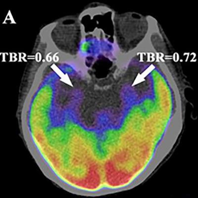
 Axial views of the amygdala. Higher amygdalar uptake of F-18 FDG in a patient who died (A) compared with a patient who survived (B) after a diagnosis of head and neck cancer. These images were recorded at the initial cancer staging prior to cancer treatment. Both individuals had the same stage and type of cancer. Image courtesy of PLOS One.
The researchers included FDG-PET/CT images from 240 patients with head and neck cancer who previously underwent FDG-PET/CT scans as part of initial cancer staging. They measured F-18 FDG uptake in the amygdala by placing circular regions of interest in the right and left amygdalae and calculated radiotracer tracer accumulation in each region.
Among the group, 67 patients died over a median follow-up period of three years. According to the analysis, individuals within the highest AmygA experienced a two-fold higher mortality rate compared to others (p = 0.01). In addition, the median progression-free survival was 25 months for patients with higher activity compared with 36.5 months in other individuals.
"AmygA, quantified on routine F-18 FDG-PET/CT images obtained at cancer staging, independently and robustly predicts mortality and cancer progression among patients with [head and neck cancer]," the group wrote.
The researchers noted that individuals in the study were aware that they were being evaluated for cancer and that this may have increased their anxiety and impacted amygdalar activity. Thus, it is unclear if AmygA on PET/CT scans would be similarly predictive of cancer outcomes in other imaging settings, they wrote.
Yet the study underscores the need for further research, especially given the well-described association between stress and cancer risk, they suggested.
"Future studies should test whether strategies that attenuate AmygA (or its downstream biological consequences) may improve cancer survival," the researchers concluded.
The full article can be found here.