
A new immuno-PET imaging method may offer a novel approach to detecting low levels of HIV in patients undergoing antiretroviral therapy (ART), according to a study published March 9 in Nature Communications.
In a first-in-human trial, a team at the University of California, San Francisco (UCSF) used PET imaging with an experimental radiotracer based on zirconium-89 (Zr-89) to detect low levels of HIV in patients undergoing ART compared with normal patients. The finding could help in efforts to eradicate the virus in HIV-positive patients.
"PET tracer uptake may be associated with tissue HIV burden and has the potential to play an important role in HIV persistence studies and viral eradication efforts," wrote corresponding author Dr. Timothy Henrich, an associate professor of medicine, and colleagues.
HIV persists in immune cells in the body despite successful suppression with antiretroviral therapy (ART). This is a major obstacle to achieving long-term free remission or a functional cure for HIV-infected patients.
Moreover, these latent reservoirs of infected cells lurk in anatomical regions that are inaccessible to routine tests. Therefore, a noninvasive method for understanding long-term HIV persistence is urgently needed, the authors wrote.
VRC01 is a monoclonal antibody designed to bind to a certain protein expressed by CD4 T cells infected by HIV. When combined with a Zr-89 radioisotope, the resulting tracer reveals levels of these "target" cells on PET scans.
In this study, the UCSF group's objective was to evaluate the distribution of Zr-89 VRC01 in lymph nodes and other tissue suspected of harboring low levels of virus in HIV-positive patients on ART, compared with uninfected individuals.
The researchers recruited 15 patients 18 years of age or older and split them into three equal groups: HIV-infected patients with high viral loads, HIV-infected patients who were on continuous suppressive ART (with viral loads < 40 RNA copies/mL prior to imaging), and uninfected patients.
PET scans were performed at four time points for all patients following the injection of the Zr-89 VRC01 tracer -- at one to two, four to six, 24, and 72 hours -- to determine the optimal timing for imaging. The researchers compared maximum standardized uptake value (SUVmax) and SUVmean levels indicating quantities in tissue of HIV-infected cells among the groups.
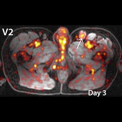
 (a) Axial images with inset magnification of bilateral inguinal lymph nodes on imaging day 3 for a viremic (V3), ART-suppressed (A1), and uninfected control (C3). (b) Axial image of persistent inguinal lymph node Zr-89 VRC01 uptake in inguinal lymph nodes on imaging day 0 (6 hours post injection), and imaging day 3 (72 hours post injection). Image courtesy of Nature Communications.
Differences in tissue uptake in HIV-positive patients compared to uninfected patients were the greatest at approximately 24 hours after Zr-89 VRC01 administration, according to the findings. The researchers observed significantly higher activity of the tracer in inguinal (groin) and axillary lymph nodes in HIV-positive patients compared to uninfected participants at all time points.
Importantly, PET tracer uptake in inguinal lymph nodes in HIV-positive patients and ART-suppressed patients significantly and positively correlated with HIV protein expression measured directly in tissue in laboratory studies, the group wrote.
"Zr-89 VRC01 uptake in various tissues (including lymph nodes and gut) is higher in HIV-infected individuals with detectable viremia and on suppressive ART compared to uninfected controls," the authors wrote.
The study represents initial data from an ongoing trial after five participants were enrolled in each of three groups (HIV on ART, HIV not on ART, and uninfected controls). The authors noted the proof-of-concept study confirmed animal studies in macaques showing that inguinal and axillary lymph nodes are major sites of HIV persistence.
Ultimately, combining PET-based imaging approaches and direct tissue studies of HIV persistence has the potential to expand our understanding of the whole-body burden and activity of HIV across the body, they wrote.
Further studies in longitudinal cohorts with direct access to a wide range of tissues are needed to fully validate the relationship between PET signal and local HIV burden, however.
"Our study provides novel immuno-PET imaging methods that may be adapted to a wide variety of studies involving low-level target expression," Henrich and colleagues concluded.


![A 53-year-old patient (patient number four) with a recurrent pituitary adenoma with extension of a cystic component of disease to the medial temporal lobe apparent on MRI (contoured in blue), and extension of disease to the left sphenoid bone and orbital apex apparent on [68Ga]Ga-DOTA-TATE (contoured in yellow).](https://img.auntminnie.com/mindful/smg/workspaces/default/uploads/2026/04/pituitary-tumor.QGsEnyB4bU.jpg?auto=format%2Ccompress&dpr=2&fit=crop&h=167&q=70&w=250)


