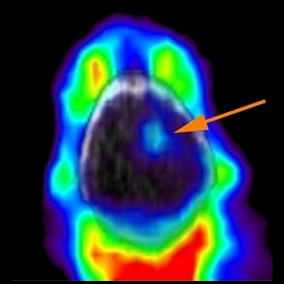
For decades, researchers have known that cancer harbors an insatiable appetite for iron. Now they may have a PET tracer, called F-18 TRX, that targets iron on cancer cells, which could help in the development of therapies that exploit iron activity in tumors.
In a laboratory and animal study published in the July issue of the Journal of Nuclear Medicine, researchers from the University of California, San Francisco (UCSF) developed a method using F-18 TRX for targeting and measuring what's known as the "labile iron pool" (LIP) in cancer cells.
"LIP levels in patient tumors have never been quantified," said UCSF pharmaceutical chemist Adam Renslo, PhD, in a news release.
Researchers imaged 10 tissue graft models of glioma and renal cell carcinoma with F-18 TRX to measure LIP. They measured the uptake of the radiotracer in the cancer. An animal model study was also conducted to determine effective human dosimetry.
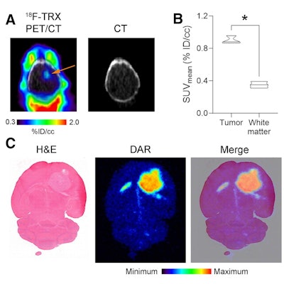
 (A) F-18 TRX PET/CT data showing radiotracer uptake in a U87 MG tumor (arrow) implanted within the right hemisphere of a mouse brain. The image was acquired at 90 minutes post injection. (B) Quantification of F-18 TRX uptake using ROI analysis of the PET data from mice bearing U87 MG tumors (n = 3). The tumor uptake was compared to uninvolved normal white matter on the contralateral region of the brain. (C) Digital autoradiography showing the distribution of the radiotracer within a coronal section of the mouse brain. The tissue was stained with H&E and merged with the pseudocolor image of the autoradiography. Image courtesy of the Journal of Nuclear Medicine
F-18 TRX showed a wide range of tumor accumulation but successfully identified LIP levels among tumors and helped determine which ones might be most likely to respond to LIP-targeted therapies. The estimated effective dose for adults was comparable to doses used with other F-18 tracers.
The research is an important milestone in understanding the therapeutic potential of LIP-targeted therapies, Renslo and colleagues stated.


