Measuring abdominal aortic calcification on routine lumbar spine x-rays can help identify elderly patients at risk for fractures, researchers have reported.
The finding is from an analysis of 1,395 hip fracture patients and 1,075 control subjects, with abdominal aortic calcification (AAC) scores of 5 or higher significantly associated with hip fractures, noted lead author Yuto Shibata, MD, of Kumamoto University in Japan, and colleagues.
"AAC score could serve as a potential surrogate marker to identify patients at potential risk of hip fracture as a means of encouraging them to take osteoporosis medications," the group wrote. The study was published June 1 in Scientific Reports.
Many patients are unaware of their fracture risk until a fracture occurs, the authors explained. Because lower back pain is highly prevalent among older adults, lumbar spine x-rays are frequently obtained in these patients. These images may provide an opportunity to assess hip fracture risk during routine evaluation, according to the researchers.
Although several studies have reported an association between AAC, a form of vascular calcification, and fracture risk, data in the Japanese population remain limited, they noted. To bridge the gap, the group analyzed 1,395 hip fracture cases (mean age 85 years old) and 1,075 non-hip fracture controls (mean age 79 years old). They assessed AAC semiquantitatively using visual scores applied to lateral lumbar spine x-rays at the L1 through L4 vertebral levels, with each segment scored up to 6 points for a maximum total of 24.
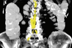
 Abdominal aortic calcification (AAC) score grading: 0 - no calcific deposits in front of the vertebrae; 1 - small scattered calcific deposits filling less than 1/3 of the longitudinal wall of the aorta; 2 - 1/3-2/3 of the wall calcified; 3 - 2/3 or more of the wall calcified.Scientific Reports
In addition, in terms of efficiency, the AAC score compared favorably with existing fracture risk tools, the researchers reported. AAC assessment on spine x-rays required approximately 10 seconds per case, versus 15.5 seconds for FRAX and more than four minutes for the K-STOP scoring system.
“Here, we show that AAC seen anteriorly on a lateral lumbar spine x-ray image could serve as a convenient tool to assess possible hip fracture risk,” the researchers wrote.
The group noted that 93% of patients with hip fractures in the study were not undergoing osteoporosis treatment at the time of fracture and had been unaware of their fracture risk. While AAC assessment requires a trained expert, it requires only about 10 seconds for a physician and offers an opportunity to raise awareness for patients, they wrote.
“The AAC assessment takes only a few seconds to complete and is not a significant burden for physicians in low back pain practice,” the group concluded.
The full study is available here.