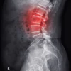
A four-year study of 200 children and adolescents with type 1 diabetes found that bone density and structure decline significantly over time, particularly in those with longer disease duration, raising concerns about reduced peak bone mass and increased fracture risk in adulthood.
- Bone mineral density Z-scores declined significantly over four years in children with type 1 diabetes, from -1.4 to -1.7
- Trabecular bone density fell from 167.7 to 150.2 mg/cm³, while cortical thickness decreased from 3.1 mm to 1.8 mm
- Adolescence is a critical period for building peak bone mass, and disruptions during this time can cause lifelong skeletal problems
- Early identification of high-risk patients and longitudinal monitoring may help preserve bone health in young diabetics
- Previous research relied mainly on cross-sectional studies, leaving a gap in long-term bone health data in low- and middle-income countries
A bone imaging study has revealed declines in bone density and structure over time in children and adolescents with type 1 diabetes.
The finding is from a retrospective, single-center analysis of 200 children and adolescents with type 1 diabetes followed for four years using dual-energy x-ray absorptiometry (DEXA) and peripheral quantitative computed tomography (pQCT), noted lead author Shruti Mondkar, MD, of the Hirabai Cowasji Jehangir Medical Research Institute in Pune, India, and colleagues.
"Skeletal health remains under-recognized, under-evaluated, and rarely followed longitudinally, despite increasing evidence of clinically significant bone deficits in this population," the group wrote. The study was published July 19 in Bone.
Childhood and adolescence are critical periods for building peak bone mass, and disruptions during this window can have lifelong consequences, including reduced peak bone mass and greater fracture risk in adulthood, the authors explained. Previous studies in children with type 1 diabetes have largely used cross-sectional designs, leaving a gap in longitudinal data on bone accrual trajectories, particularly in low- and middle-income settings, the authors noted.
To address the gap, the researchers analyzed records from 200 participants in the Sweetlings cohort, a structured longitudinal diabetes care program at a tertiary care hospital in Western Maharashtra, India, collected between December 2015 and December 2022. Participants had a mean age of 10.6 years. Among the subjects, 160 had type 1 diabetes for under five years and 40 for five years or more.
The DEXA analysis showed total body less head bone mineral density (BMD) Z-scores declined significantly over four years (-1.4 ± 1.1 to -1.7 ± 1.2), while lumbar spine bone mineral apparent density Z-scores remained unchanged.
On pQCT, trabecular volumetric BMD fell (167.7 ± 35.6 to 150.2 ± 39.8 mg/cm³), as did trabecular density-for-age Z-scores (-0.9 ± 1.5 to -1.7 ± 1.2) and total density-for-age Z-scores (-0.5 ± 1.5 to -1.0 ± 1.0). Cortical volumetric BMD increased over the same period, but cortical thickness declined significantly (3.1 ± 0.6 mm to 1.8 ± 0.3 mm), the authors reported.
“Children and adolescents with [type 1 diabetes] demonstrate progressive deterioration in bone health over time, particularly those with longer disease duration and adverse metabolic profiles,” the group wrote.
Ultimately, early identification of high-risk individuals and longitudinal monitoring may help preserve bone health in these patients, the group suggested.
However, further prospective studies incorporating healthy controls and clinically relevant outcomes, including fracture risk, are needed before these findings can inform screening or management recommendations, the researchers concluded.
The full study is available here.