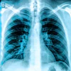
The first x-rays taken of humans in space demonstrated diagnostic quality equivalent to preflight images, according to a study published in Radiology. Three crew members with only four hours of training successfully acquired anatomic x-rays during the Fram2 mission, proving that portable x-ray imaging is feasible in actual orbital spaceflight and expanding diagnostic capabilities for crew health monitoring.
- Diagnostic Equivalence: In-flight x-rays showed no significant differences in overall image quality, spatial resolution, or contrast resolution compared to preflight images (p > .99).
- Crew Training: Three nonmedical crew members with approximately four hours of training successfully performed the imaging procedures in microgravity.
- Study Scope: The Fram2 mission lasted 3.5 days and included x-rays of hands, forearms, chest, abdomen, and pelvis.
- Primary Challenge: Positioning of central body images (chest, pelvis, abdomen) was worse in-flight than preflight, identified as the main operational difficulty.
- Future Steps: Researchers recommend standardizing microgravity protocols, developing lightweight mounting systems, and integrating real-time acquisition support for next-generation space imaging.
The first x-rays taken of humans in space match the overall image quality of those taken preflight, according to research published July 14 in Radiology.
The finding is from a prospective feasibility study of three crewmembers during the 3.5-day Fram2 polar orbital flight, in which crew members acquired anatomic x-rays without live ground support, noted lead author Sheyna Gifford, MD, of the Mayo Clinic in Rochester, MN, and colleagues.
“While central body images (chest, pelvis, and abdomen) had worse positioning in-flight, in-flight radiographs were equivalent to preflight radiographs in overall image quality, spatial resolution, and contrast resolution,” the group wrote.
While earlier work showed that portable x-ray was feasible during simulated microgravity conditions, no system had previously been tested during actual orbital spaceflight, the authors noted. During the Fram2 mission, three crewmembers (mean age, 42.8 years; two women) who had received about four hours of training acquired anatomic radiographs of the hand, forearm, chest, abdomen, and pelvis, both preflight and in-flight.
For the study, the x-rays were evaluated by independent radiologists on overall image quality, spatial resolution, contrast resolution, and positioning (Likert scores of 1–5). Secondly, preflight x-rays were compared with in-flight x-rays using the Wilcoxon rank sum test, while crew surveys were conducted postflight.
 Representative preflight, in-flight, and postflight chest radiographs. Radiographs of the chest were acquired (A) preflight by a crewmember, (B, C) in-flight on day 3 after launch (L+3) by a crewmember, and (D) postflight by a non-crew operator using the same imaging protocol.RSNA
Crew surveys identified alignment and positioning, including of the subject, x-ray source, and detector, as the primary operational challenges, while equipment and protocols were rated easy to use.
“Our study demonstrates the feasibility of in-orbit radiography and expanded diagnostic capabilities for crew health and hardware evaluation,” Gifford said, in a news release from RSNA. “Acquiring diagnostically useful x-rays in space is something that anyone can do. Three very talented nonmedical people with four hours of training in one of the harshest environments did it right and did it well.”
In an accompanying editorial, Suhny Abbara, MD, of Mayo Clinic in Jacksonville, FL, and Alan B. McMillan, PhD, of the University of Wisconsin-Madison, noted that as an early feasibility study, the work necessarily has limitations, including small sample size, short mission duration, and constrained imaging scenarios.
“But these limitations clarify the path forward,” the pair wrote.
The major next steps are standardization of microgravity acquisition protocols, development of lightweight mounting and alignment systems, and integration of real-time acquisition support, Abbara and McMillan concluded.
Read the full study here.