Dual-energy x-ray absorptiometry (DEXA) exams may offer a novel approach for identifying older women at risk of dementia, according to a study published June 26 in the Lancet Regional Health -- Western Pacific.
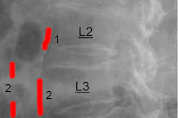
Using images acquired in a large group of older women in 1998, researchers in Australia analyzed DEXA lateral spine images for signs of abdominal aortic calcification (AAC) -- an early marker associated with vascular disease. Fourteen years after the images were acquired, the researchers found that women who had high levels of AAC were twice as likely to be hospitalized or die from dementia.
"These scans may be a cheap, rapid, and safe way to screen a large number of susceptible older Australians for higher late-life dementia risk," said co-author Simon Laws, PhD, of Edith Cowan University in Joondalup, in a news release from the university.
Late-life dementia -- occurring after the age of 80 -- is increasingly common due to vascular and nonvascular risk factors. Therefore identifying individuals who are at higher risk of dementia remains a global priority.
DEXA bone density testing is widely used in osteoporosis screening of older men and women. DEXA lateral spine imaging can easily and rapidly also capture AAC levels, which are known to be associated with early coronary artery plaque deposits (atherosclerosis) and thus poor long-term health outcomes in older men and women.
To date, just two studies in middle-aged and older men and women have found that the presence and extent of AAC are associated with poorer cognitive function, and no prior studies have investigated associations between AAC and dementia risk, the authors wrote.
In this work, the researchers analyzed AAC levels on DEXA lateral spine images acquired in 958 older women (averaging 75 years old) who participated in an osteoporosis study in 1998. At baseline, 44.7% of the women had low AAC, 36.4% had moderate AAC, and 18.9% had extensive AAC.
Over more than 14 years, 150 (15.7%) women experienced hospitalization from late-life dementia (n = 132) and/or death (n = 58).
An analysis revealed that compared with women with low AAC, women with moderate and extensive AAC were more likely to suffer late-life dementia hospitalization and death. After adjusting for other cardiovascular and genetic risk factors for Alzheimer's disease, women with moderate and extensive AAC had twice the relative hazard ratio (HR) of late-life dementia (moderate, aHR = 2.03; extensive, aHR = 2.10) compared with women with low AAC.
"We demonstrated that over half of the older women with AAC were at two to four times the risk of late-life dementia hospitalization and deaths over the next 15 years," the authors wrote.
This is the first study to report an association between AAC and the risk of future late-life dementia, and it will be important to replicate these findings in other cohorts, the authors noted.
Ultimately, the researchers suggest that assessing AAC at the time of DEXA bone density testing could provide a window of opportunity to enable dietary, lifestyle, and cardiovascular risk management modifications to prevent the progression of dementia in older women.
With some 600,000 bone density tests performed each year in Australia to screen for osteoporosis, the researchers are now working with a computer science team to automate AAC assessments on DEXA images, which could make the process quicker and easier, they noted.
"I think the next step is telling people about their AAC and late-life dementia risk to see if this can motivate healthy diet and lifestyle behavior changes," Laws concluded.