Doctors may be able to distinguish between calcified and noncalcified lung nodules on patient chest x-rays using a one-shot dual-energy subtraction (DES) technique, a group from Niigata University in Japan has found.
A team led by Motohiko Yamazaki, MD, compared soft tissue images obtained by a one-shot DES method compared with standard images alone and found the accuracy of five radiologists improved using the experimental technique.
"The soft tissue images obtained using one-shot DES with a flat-panel detector have added value in distinguishing calcified from noncalcified nodules on chest radiographs, especially for less experienced radiologists," the group wrote.
Calcification is one of the most reliable indicators of whether or not solitary pulmonary nodules are benign. The accuracy of standard chest x-rays for detecting calcification is low, however, as small nodules may be obscured by overlapping structures such as the ribs, the authors explained.
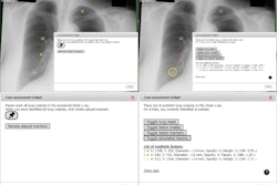
 A case of a calcified nodule overlapping with the bone. On the standard image (left), the presence or absence of calcification among five readers was not consistent (confidence level of each reader: 4, 3, 3, 4 and 2). In the soft tissue image (right), the nodule disappeared. All readers correctly evaluated it as calcified (confidence level of each reader: 1, 1, 2, 2 and 2). Image and caption courtesy of Scientific Reports through CC BY 4.0.
DES is a technique based on acquiring two separate images from patients -- a low-energy and high-energy x-ray image -- which are then processed with structures that potentially obscure nodules in soft tissue subtracted from the reconstructed images. The recent development of flat panel digital detectors that can absorb both the low-energy and high-energy x-ray beams simultaneously has made a "single-shot" DES approach more feasible, they noted.
To evaluate the technique, the investigators identified 139 patients in whom both standard x-rays and one-shot DES images were acquired by the same system. The images included 155 nodules, of which 48 were calcified and 107 noncalcified. Five radiologists then interpreted whether or not the nodules were calcified using standard x-rays and then DES x-ray images.
According to the analysis, the accuracy of all radiologists increased with the addition of soft tissue one-shot DES images.
| Accuracy among readers detecting nodule calcification on standard and one-shot DES x-rays | ||
| Readers (years of experience) | Standard x-ray | DES soft tissue x-ray |
| Reader 1 (26 years) | 89.7% | 92.3% (p = 0.206) |
| Reader 2 (14 years) | 83.2% | 87.7% (p = 0.178) |
| Reader 3 (8 years) | 79.4% | 92.3% (p < 0.001) |
| Reader 4 (6 years) | 77.4% | 87.1% (p = 0.007) |
| Reader 5 (3 years) | 63.2% | 83.2% (p < 0.001) |
"We showed that the use of standard images along with soft tissue images improved the distinguishability between calcified and noncalcified nodules, particularly for less experienced radiologists," the researchers stated.
The one-shot DES approach solves the problem of aligning two separate x-rays images acquired using dual-exposure DES, which involves adjusting for patient movement at different time points, the authors noted.
Nonetheless, more research is warranted, they noted.
"To the best of our knowledge, the present study is the first to evaluate the effectiveness of one-shot DES with a flat-panel detector to differentiate between calcified and noncalcified nodules," the group concluded.
The article was published June 12 in Scientific Reports.