Researchers at Semmelweis University's Medical Imaging Centre in Budapest, Hungary used a photon-counting CT (PCCT) scanner to examine Egyptian mummy remains from the Semmelweis Museum of Medical History.
The exam, conducted outside patient hours in accordance with standard clinical practice, covered multiple specimens including two mummified heads, two lower limb remains, a foot, and a mummified hand. The oldest remains have been dated to between 401 and 259 BCE, making them more than 2,300 years old, the researchers said.
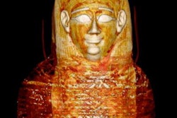
 Researchers in Hungary used PCCT imaging to examine Egyptian mummy remains, such as this mummified head.Medical Imaging Center (OKK), MNMKK Semmelweis Museum of Medical History
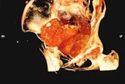
Among the findings, the new images allowed researchers to identify possible osteoporosis in a previously examined lower limb, for which no definitive diagnosis had been possible using earlier CT scans. High-resolution imaging of the two mummified heads is expected to support more accurate age determination and lay the groundwork for future 3D and facial reconstructions, the researchers said.
One specimen previously thought to be a human head or bird mummy was confirmed by an earlier CT scan to be an adult foot, and the new images are being used to analyze the mummification technique and bandage layers in greater detail.
The university said a detailed evaluation of the images is currently underway.