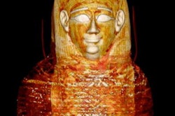
Remember the Pa-Ib (pronounced pie-eeeb) mummy -- also known as "Ipy's mummy" -- on which researchers used 64-slice CT imaging to uncover its secrets? It now has its own documentary.
Produced by the Barnum Museum in Bridgeport, CT, the 30-minute documentary, titled "Uncovering the Secrets of an Egyptian Mummy and Coffin: The Quest to Restore Personhood," gives an in-depth look at the mummy's history.
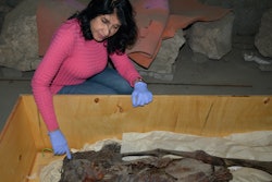
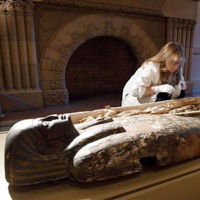
 The Barnum Museum has released a documentary on the history of a mummy on which researchers used CT imaging. Image courtesy of the Barnum Museum.
Along with investigating the transcriptions on the mummy's coffin, the documentarians also feature CT scans acquired by Gerald Conlogue from Quinnipiac University that were featured in National Geographic's "Mummy Roadshow." The museum said these scans provided the necessary data for facial reconstruction.
The documentary can be found on the Barnum Museum's YouTube page.